Figure 2. Still-shots of an RPE flatmount dissection. A: The smooth surface of an eye globe after removal of fat and extra-ocular muscles. B: Corneal penetration with a stab knife as forceps steady the eye. C: Scissors insertion at the corneal puncture cutting along the radial axis toward the optic nerve. D: The flattened globe, with neural retina remaining, after four radial cuts. E: Neural retina removal exposes the RPE for subsequent staining.
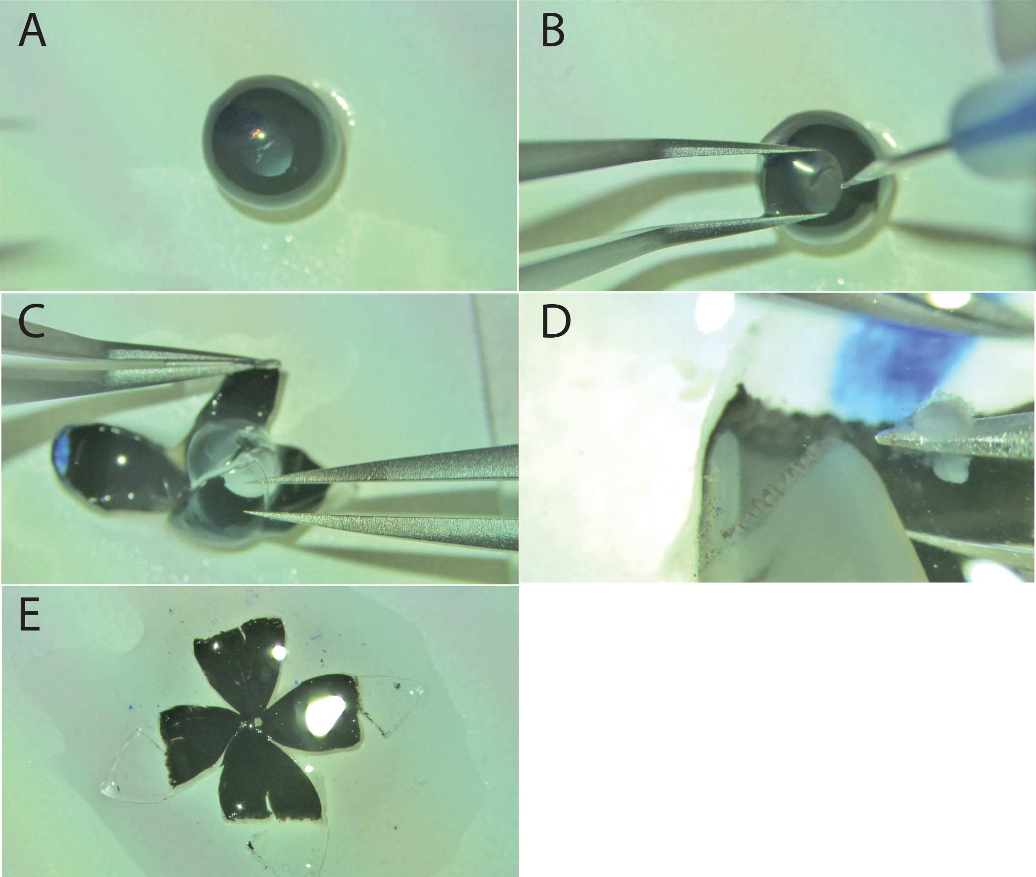
 Figure 2 of
Boatright, Mol Vis 2015; 21:40-60.
Figure 2 of
Boatright, Mol Vis 2015; 21:40-60.