Figure 17. Strain differences in eccentricity. Three genotypes, one wild type (WT), C57Bl/6J, and two models of retinal degeneration,
IRBP−/− and rd8, at postnatal day (P)30 and P720 were compared. Group sizes were four to five animals for each genotype at P30 and three
animals for each genotype at P720. The rd8 was slightly reduced in average eccentricity at P30 compared to WT, but there was no difference in eccentricity between IRBP−/− and WT at P30. As the WT aged, its RPE cells retained the same eccentricity. There was a slight increase in eccentricity
in the IRBP−/− between P30 and P720. However, the rd8 showed markedly increased average eccentricity at P720 compared to P30. Additionally, the IRBP−/− was slightly more eccentric than the age-matched WT. There was a large difference between the rd8 and the WT at P720.
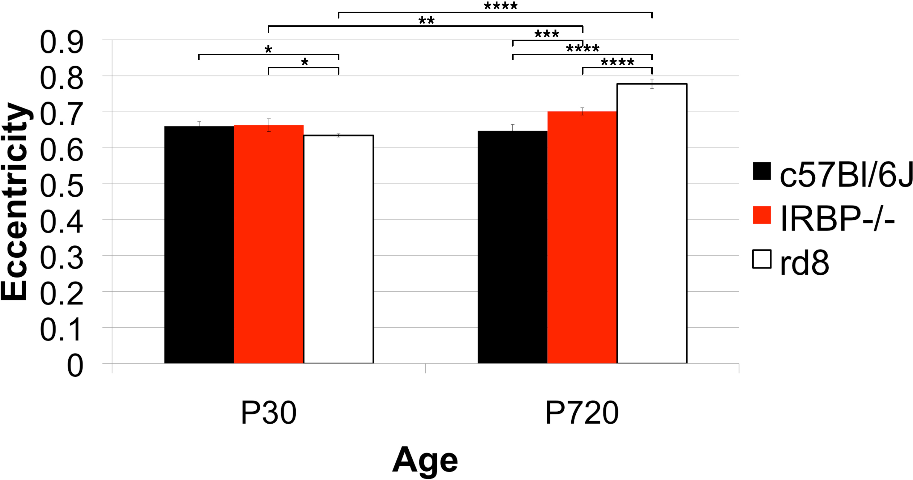
 Figure 17 of
Boatright, Mol Vis 2015; 21:40-60.
Figure 17 of
Boatright, Mol Vis 2015; 21:40-60.