Figure 1. Immunocytochemistry of RPE cells indicating RPE cell identity and culture purity. The immunostaining procedure was performed
as described in the Methods section. To confirm the epithelial origin of the cultures, cytokeratin 8/18 expression was assessed,
and to confirm that isolated cells were RPE cells, RPE65, which is involved in converting all-trans retinol to 11-cis retinal during phototransduction,
was surveyed. A: Nuclei stained blue with 4,6-diamidino-2-phenyindole dihydrochloride (DAPI). B: RPE cells stained positively for the fluorescein isothiocyanate (FITC)-conjugated cytokeratin antibody (green). C: Merged image (FITC-labeled cytokeratin and DAPI). D: DAPI-stained RPE cell nuclei (blue). E: RPE cells stained positively for the RPE65 antibody (green). F: Merged image (FITC-labeled RPE65 and DAPI; 400X).
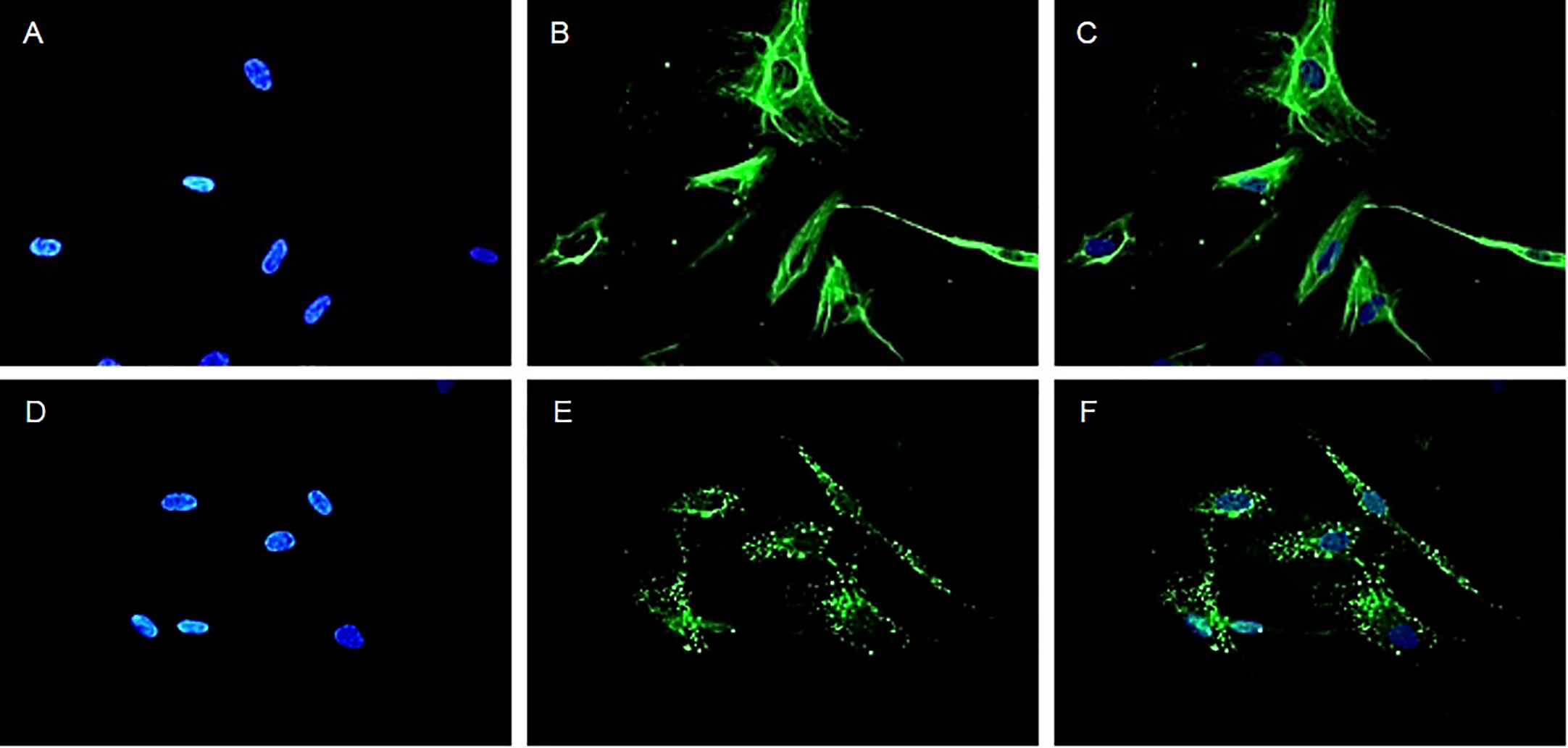
 Figure 1 of
Bagheri, Mol Vis 2015; 21:378-390.
Figure 1 of
Bagheri, Mol Vis 2015; 21:378-390.