Figure 4. Effect of hBest1 and hBest1W93C expression on Ca2+-stimulated changes in transepithelial electrical properties. A: fhRPE cells were treated with 500 nM ionomycin, which caused a biphasic increase in TEP. Each phase of the response was
measured at the indicated point. When the TEP reached a plateau, 100 µM NFA was added to the bathing solution to inhibit calcium-activated
anion channel activity. Representative recordings of responses are shown in (B), demonstrating that overexpression of either hBest1 or hBest1W93C resulted in a suppression of the overall response, and that the response to the addition of NFA was similar in control and
monolayers overexpressing hBest1. Isc (C) was determined from TEP (D) and TER (E) for P0, P1, P2, and NFA. Responses were elevated at P0 for hBest1-overexpressing monolayers compared to controls and monolayers
overexpressing hBest1W93C. However, at P1, P2, and NFA, Isc was not significantly different between controls and monolayers overexpressing hBest1. Neither ionomycin nor NFA had a significant
effect on the transepithelial electrical properties of fhRPE monolayers expressing hBest1W93C. Compared to control and hBest1-overexpressing monolayers, TEP (D) and Isc (C) were significantly reduced for hBest1W93C monolayers at P0, P1, P2, and NFA. For both control and hBest1-overexpressing monolayers, the differences in TEP (D) and Isc (C) between different time points were significant (p<0.05). Data are mean ± SD, n=9 for control, n=5 for hBest1, and n=6 for
W93C.
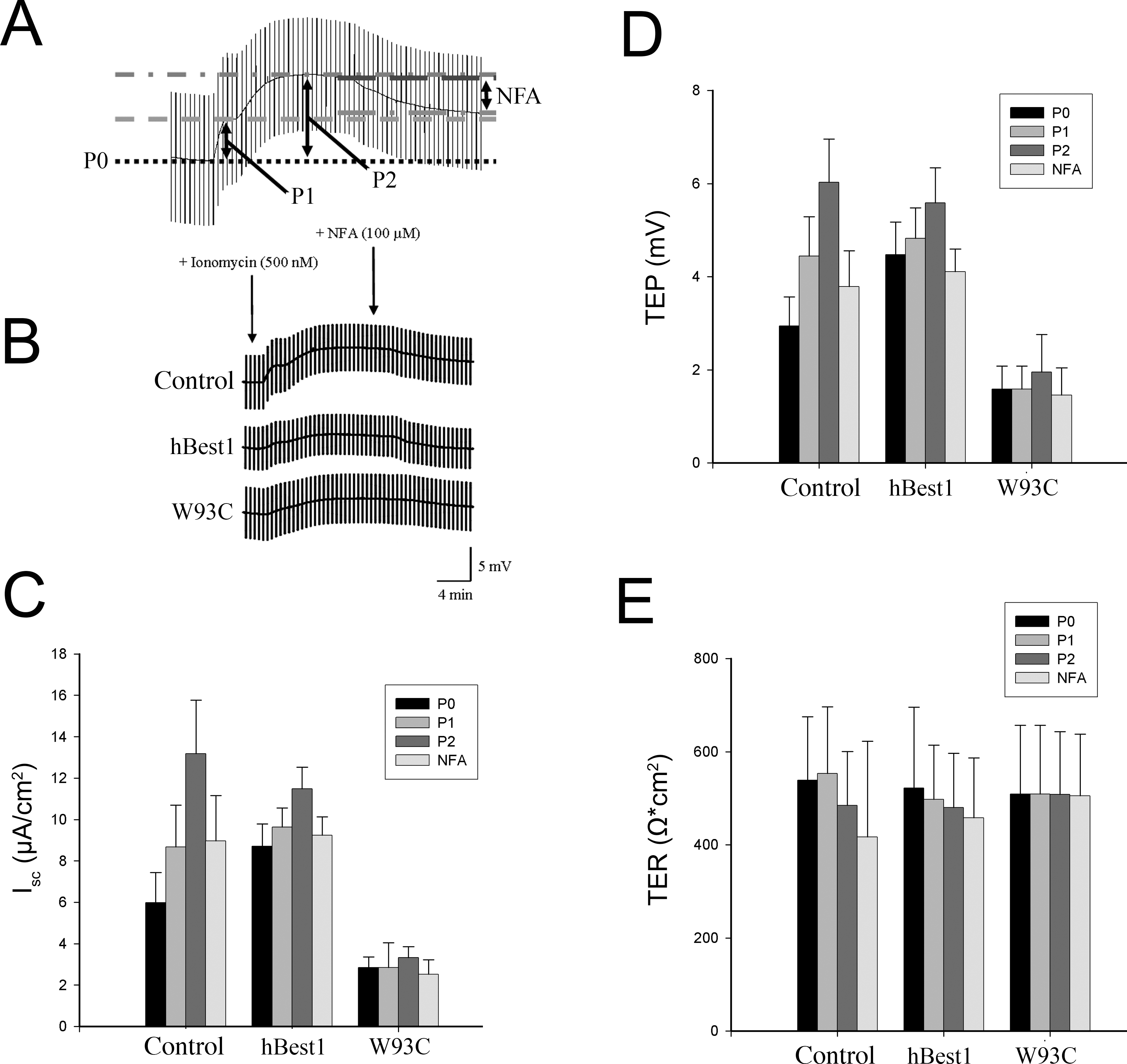
 Figure 4 of
Marmorstein, Mol Vis 2015; 21:347-359.
Figure 4 of
Marmorstein, Mol Vis 2015; 21:347-359.