Figure 2. Retinal imaging findings. A: Funduscopy revealed the salt-and-pepper appearance of the retina, characteristic of juvenile retinitis pigmentosa (RP).
B: Optical coherence tomography (OCT) image of the right eye. C: OCT image of the left eye. OCT imaging showed an overall decrease in retinal thickness with shortening of the photoreceptor
outer segments, reduction in the outer nuclear layer, and RPE atrophy. The foveal photoreceptor layer was more preserved.
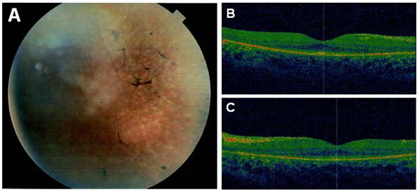
 Figure 2 of
Mayer, Mol Vis 2015; 21:306-315.
Figure 2 of
Mayer, Mol Vis 2015; 21:306-315.