Figure 1. Fundus photographs and fluorescein angiographs of two cases carrying the Arg1210Cys variation. Case 1 displayed numerous small
drusen (arrow) in the posterior pole and in the peripheral retina in both eyes. A, B: Color fundus photographs of the posterior pole and the periphery of the left eye, respectively, of case 1. C, D: Fluorescein angiographs of the posterior pole and the periphery of the left eye, respectively, of case 1. Case 2 showed
small drusen of the posterior pole in both eyes. E, F: Color fundus photographs of the right and left eyes, respectively, of case 2. G, H: Fluorescein angiographs of the right and left eyes, respectively, of case 2. In addition, case 2 displayed a large fibrotic
scar (G, arrow) in the right eye. In the left eye, pigmentary changes (F, arrow) and an occult choroidal neovascularization (H, arrow) were observed.
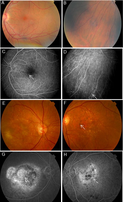
 Figure 1 of
Duvvari, Mol Vis 2015; 21:285-292.
Figure 1 of
Duvvari, Mol Vis 2015; 21:285-292.