Figure 5. Ultrastructural analysis of RPE and sub-RPE areas from Wtwt/wt and Ctrp5+/− ;wt/wt mice. Areas enclosed in boxes in A–D are magnified in panels E–H. RPE from control mice (A, E) showed no abnormalities at 21 months of age. The RPE basal infoldings are organized (star), the cytoplasm is homogenous,
and Bruch’s membrane is intact (bracket). In contrast, the RPE of the Ctrp5+/−,wt/wt mice (B, C, D, F, G, H) has structural aberrations beginning at 9 months of age. The RPE basal infoldings are disorganized (stars). Numerous packets
of undigested membranous debris are found in the basal RPE (black arrowheads). Multiple vacuoles (white arrows) and phagolysosomes
(black arrows) are found throughout the cytoplasm. Focal basal laminar deposits (white arrowheads) are present in Bruch’s
membrane, as are basal linear deposits (asterisks). All marking are identical to those used in all panels. Magnification bars=200
μm in the top row and 500 μm in the bottom row.
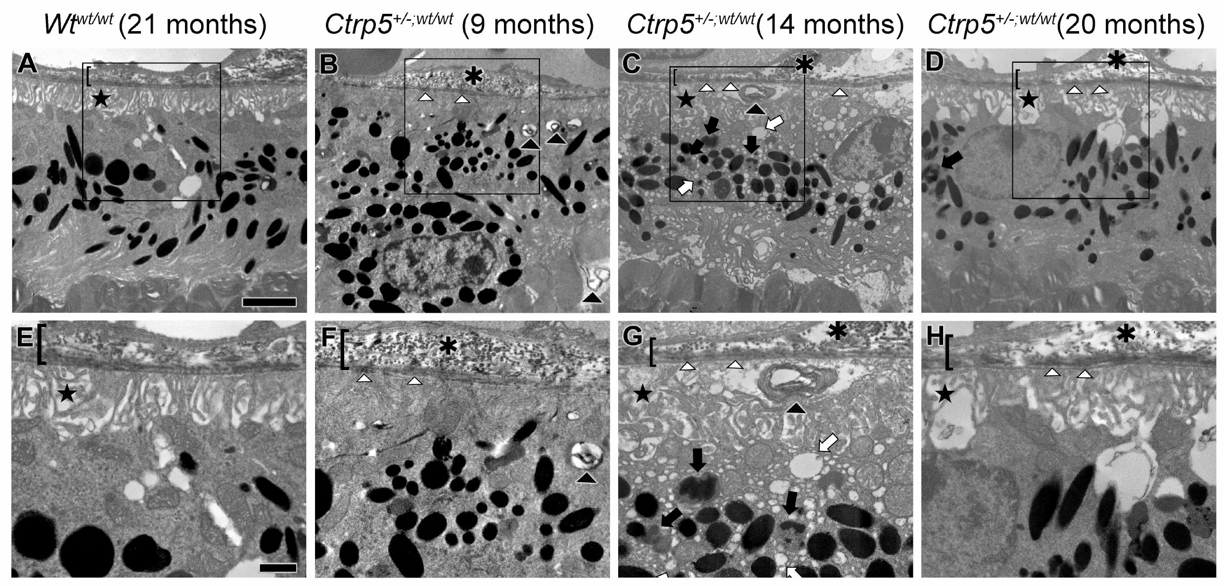
 Figure 5 of
Sahu, Mol Vis 2015; 21:273-284.
Figure 5 of
Sahu, Mol Vis 2015; 21:273-284.