Figure 3. Histological analysis of the retina structure of 8-month-old Wtwt/wt and Wtrd8/rd8 mice. The retinas from 8-month-old Wtwt/wt and Wtrd8/rd8 mice showed no dysplasia. mo, month; RPE, retinal pigment epithelium; OS, outer segment; IS, inner segment; ELM, external
limiting membrane; ONL, outer- nuclear layer; OPL, outer plexiform layer; INL, inner nuclear layer; IPL, inner plexiform layer;
GCL, glial cell layer. Magnification bar=2 μm.
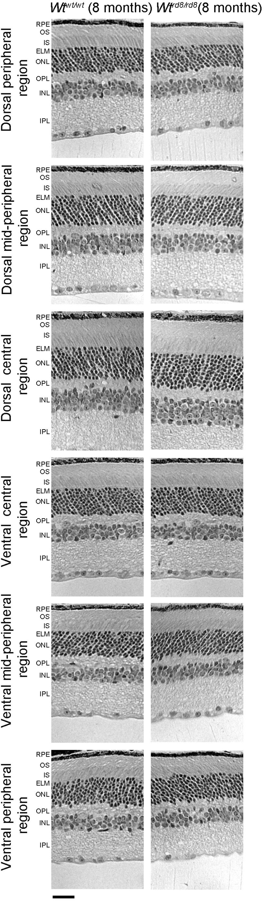
 Figure 3 of
Sahu, Mol Vis 2015; 21:273-284.
Figure 3 of
Sahu, Mol Vis 2015; 21:273-284.