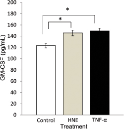
Figure 4. The RPE secreted higher levels of GM-CSF after stimulation with HNE and TNF-α. The GM-CSF secreted into the culture supernatant
was increased when primary RPE cells were exposed to 4-hydroxynonenal (HNE), an agent that promotes oxidative stress, at 10
µM for 6 h (mean ± SEM, 145.88±5.06 pg/ml versus 123.27±4.05 pg/ml, n=3, Student t test, *p<0.05). In addition, RPE cells stimulated with 20 ng/ml tumor necrosis factor alpha (TNF-α) for 6 h also resulted
in increased levels of GM-CSF secreted into the culture supernatant (149.32±3.76 pg/ml versus 123.27±4.05 pg/ml, n=3, Student
t test, *p<0.05).

 Figure 4 of
Wang, Mol Vis 2015; 21:264-272.
  Figure 4 of
Wang, Mol Vis 2015; 21:264-272.