Figure 3. Genetic analysis of C8orf37. Sanger sequencing chromatograms depicting mutations c.244–2A>C in an affected individual (48–3) from family MA48 (A) and c.555G>A; p.W185* in an affected individual (863) from family MA13 (B). Chromatograms from a normal control subject are also shown for comparison. Diagrammatic representation of the minigene
splicing assay (C) and sequencing chromatograms (D) showing that the consequence of the c.244–2A>C splice site mutation was activation of a cryptic splice site within exon
3 and deletion of 22 base pairs of coding sequence in the resulting transcript. The wild-type minigene assay spliced exons
2 and 3 correctly.
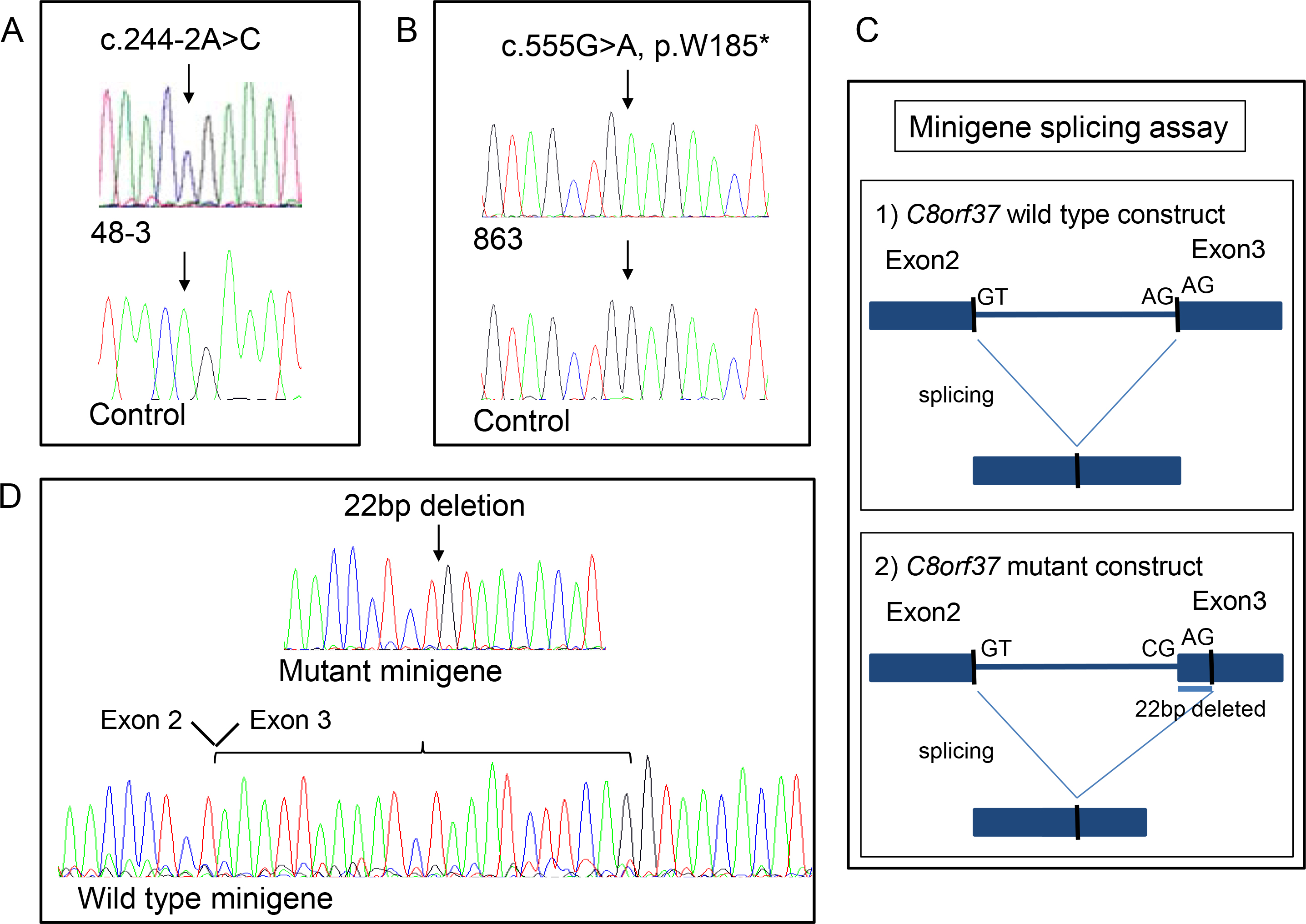
 Figure 3 of
Ravesh, Mol Vis 2015; 21:236-243.
Figure 3 of
Ravesh, Mol Vis 2015; 21:236-243.