Figure 2. Fundus photographs of patient MA48–2 at 33 years old. Color (A) and black-and-white (B) photos of the right eye show diffuse nummular hyper-pigmentation at the macula (arrows), waxy pallor of the optic disc,
arteriolar attenuation, and chorioretinal atrophy. C: Fluorescein angiography of the left eye shows bone spicule-like deposits as fluorescent areas nasal to the optic disc (arrows).
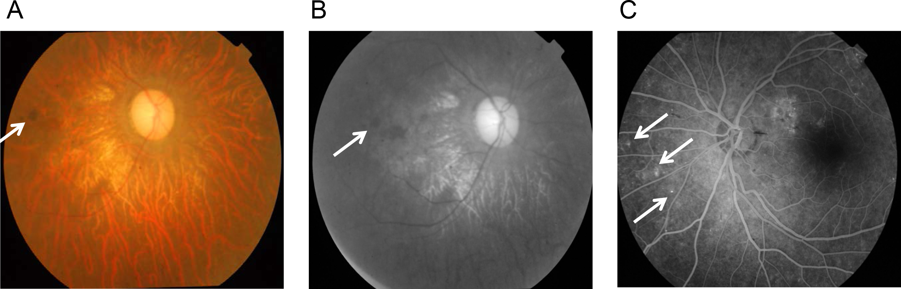
 Figure 2 of
Ravesh, Mol Vis 2015; 21:236-243.
Figure 2 of
Ravesh, Mol Vis 2015; 21:236-243.