Figure 3. Fundus photography results for the right eyes of the probands carrying the c.626G>C, c.121delG, and c.529–530delGCinsAT variants
and of a normal control. Typical changes that occur due to high myopia, including the “tigroid” appearance of the posterior
retina, are shown; the optic nerve head crescent is shown in all of the fundus photographs of the probands.
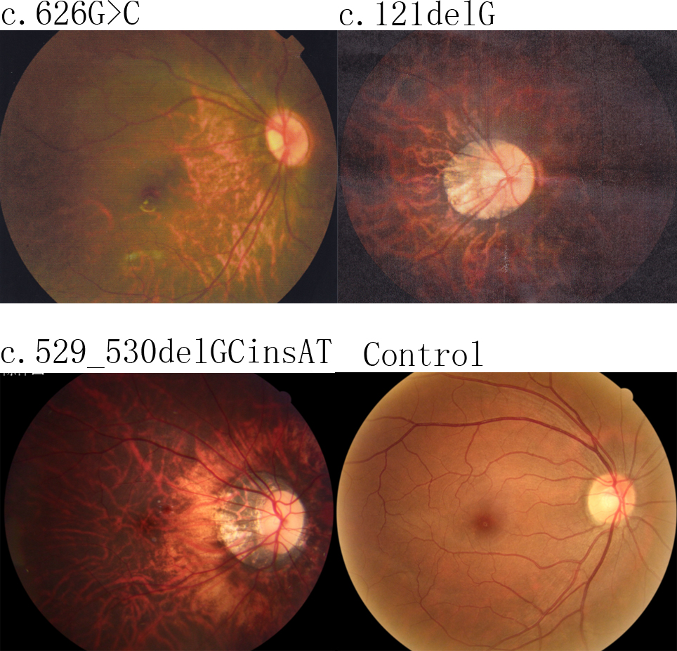
 Figure 3 of
Zhou, Mol Vis 2015; 21:213-223.
Figure 3 of
Zhou, Mol Vis 2015; 21:213-223.