Figure 1. Western blots of ABCB1, ABCC1, TLR4, and CYP3A5. Cell lysates (10 µg protein load) of RAW 264.7 mouse monocyte cells (RAW),
rat pheochromocytoma cells (PC12), and trabecular meshwork (TM) cells were resolved with sodium dodecyl sulfate–polyacrylamide
gel electrophoresis (SDS–PAGE). β-actin (10 µg protein load) was used as a loading control for each cell lysate. Data represent
a representative western blot of replicate samples.
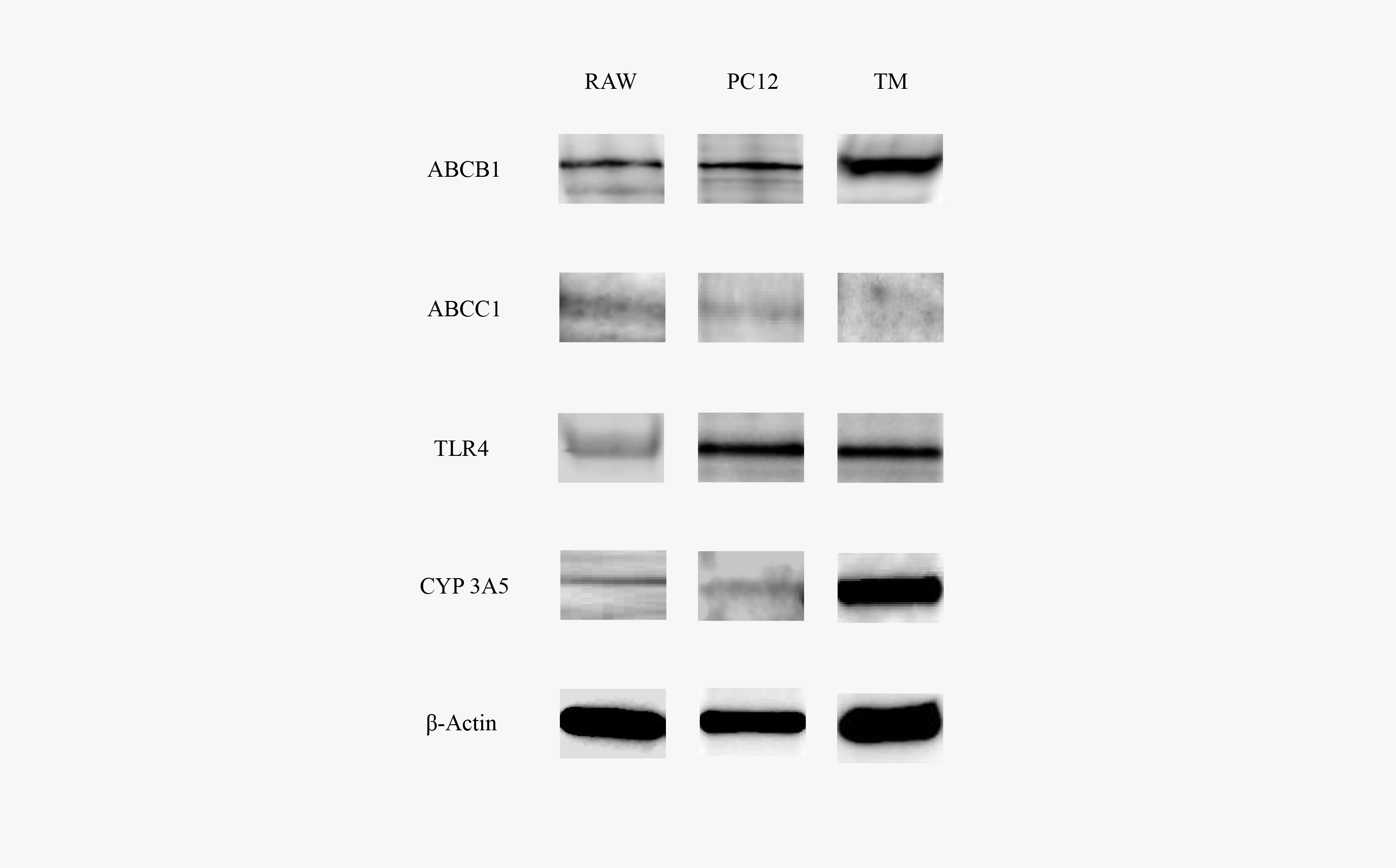
 Figure 1 of
Grybauskas, Mol Vis 2015; 21:201-212.
Figure 1 of
Grybauskas, Mol Vis 2015; 21:201-212.