Figure 4. In vitro and in vivo electroporation of sh-Kcnj10 in postnatal retina. A-D: Control or Kcnj10 expressing plasmids with enhanced green fluorescent protein (EGFP) expression plasmid were electroporated
into the isolated retina at P1 (in vitro electroporation) or the retina of live mice at P1 (in vivo electroporation). Then,
the retinas were harvested (in vitro samples), or the mice were euthanized, and the retinas were isolated (in vivo samples),
and the EGFP and retinal markers were immunostained. B: The subretinal distribution of EGFP-positive cells. In the sh-Kcnj10 sample, the outer nuclear layer (ONL) was horizontally
divided equally, and the number of cells was counted in each area (the outer half and the inner half of the ONL). C: Clonal size was counted by the number of EGFP-positive cells in vertically the same column. A and C show immunostaining pattern, and nuclei were visualized with4',6-diamidino-2-phenylindole (DAPI) staining.
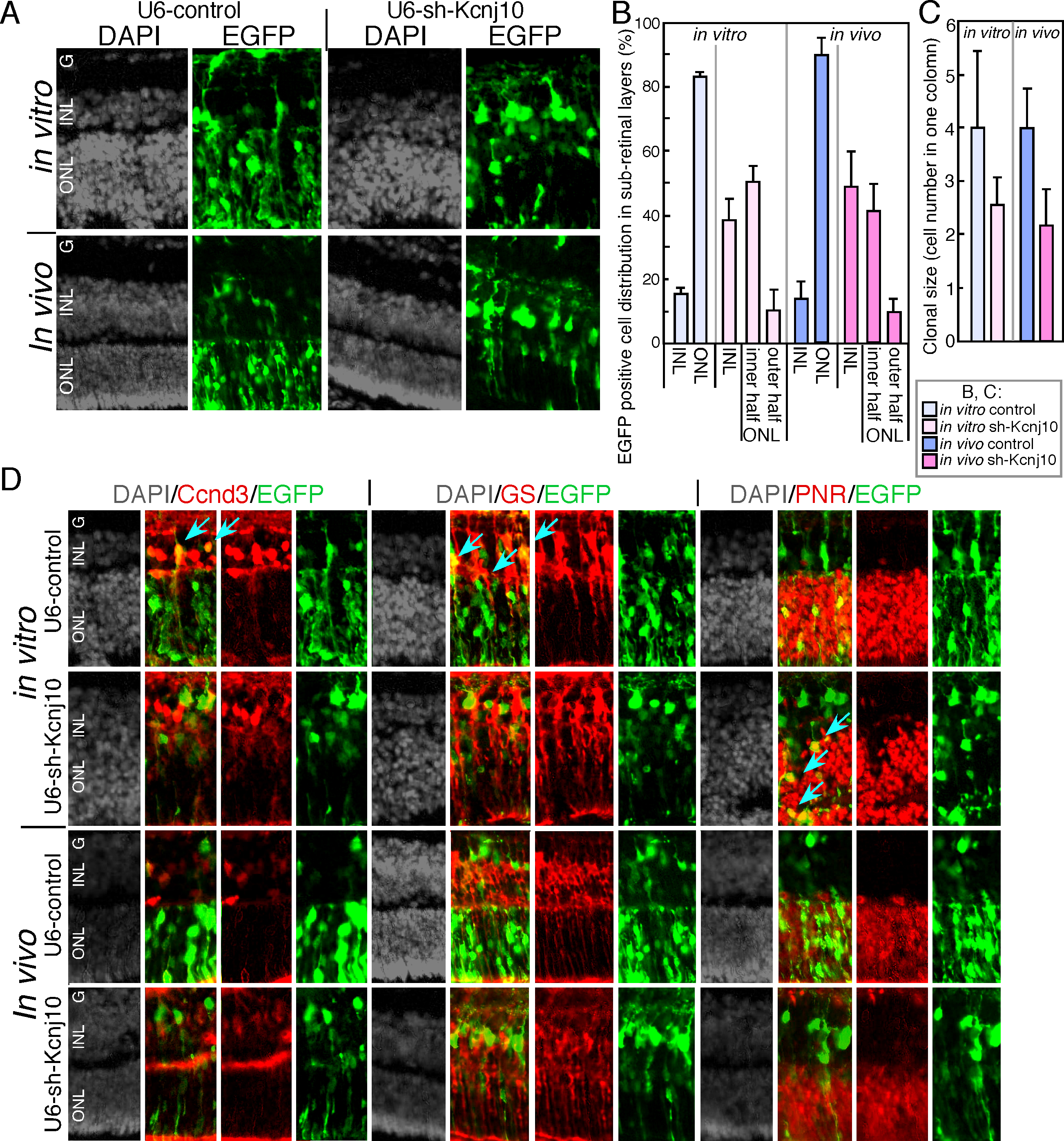
 Figure 4 of
Arai, Mol Vis 2015; 21:148-159.
Figure 4 of
Arai, Mol Vis 2015; 21:148-159.