Figure 4. Expressions of the α-SMA and Col2 proteins in the 3-D pellets of SSPCs, as stimulated by TGF-β2. In a culture medium supplemented
with 10 ng/ml of TGF-β2, the 3-D pellets grew during the 4-week culturing period. Protein expressions and localization were
visualized by a DAB reagent. (A) The negative control without the Col2 antibody. (B) Col2 expressed in the local, mid-peripheral areas of the TM pellets (arrow). (C) The negative control without the α-SMA antibody. (D) The α-SMA expression was more extended within the TM pellets, especially in the mid-peripheral and peripheral areas (arrow).
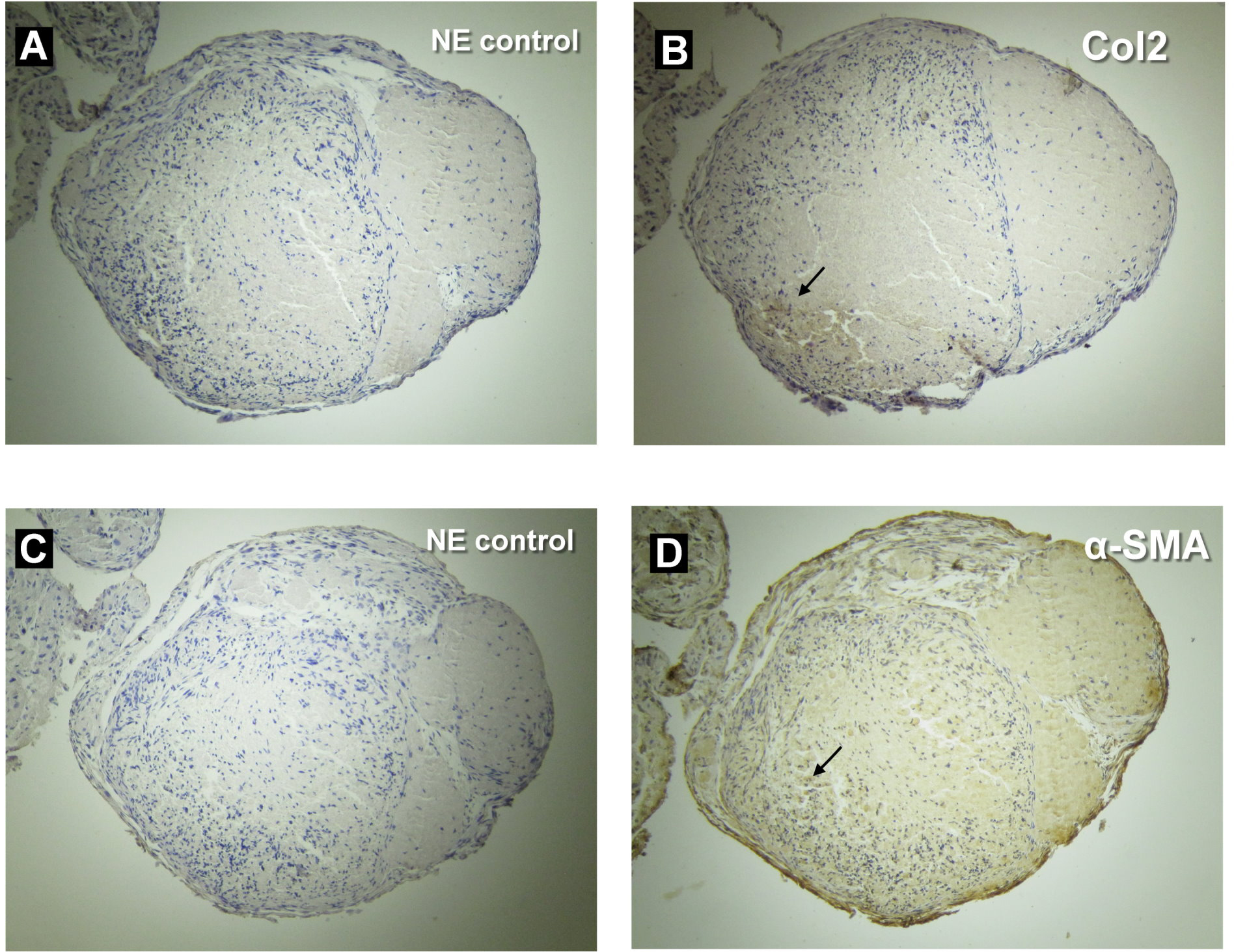
 Figure 4 of
Wu, Mol Vis 2015; 21:138-147.
Figure 4 of
Wu, Mol Vis 2015; 21:138-147.