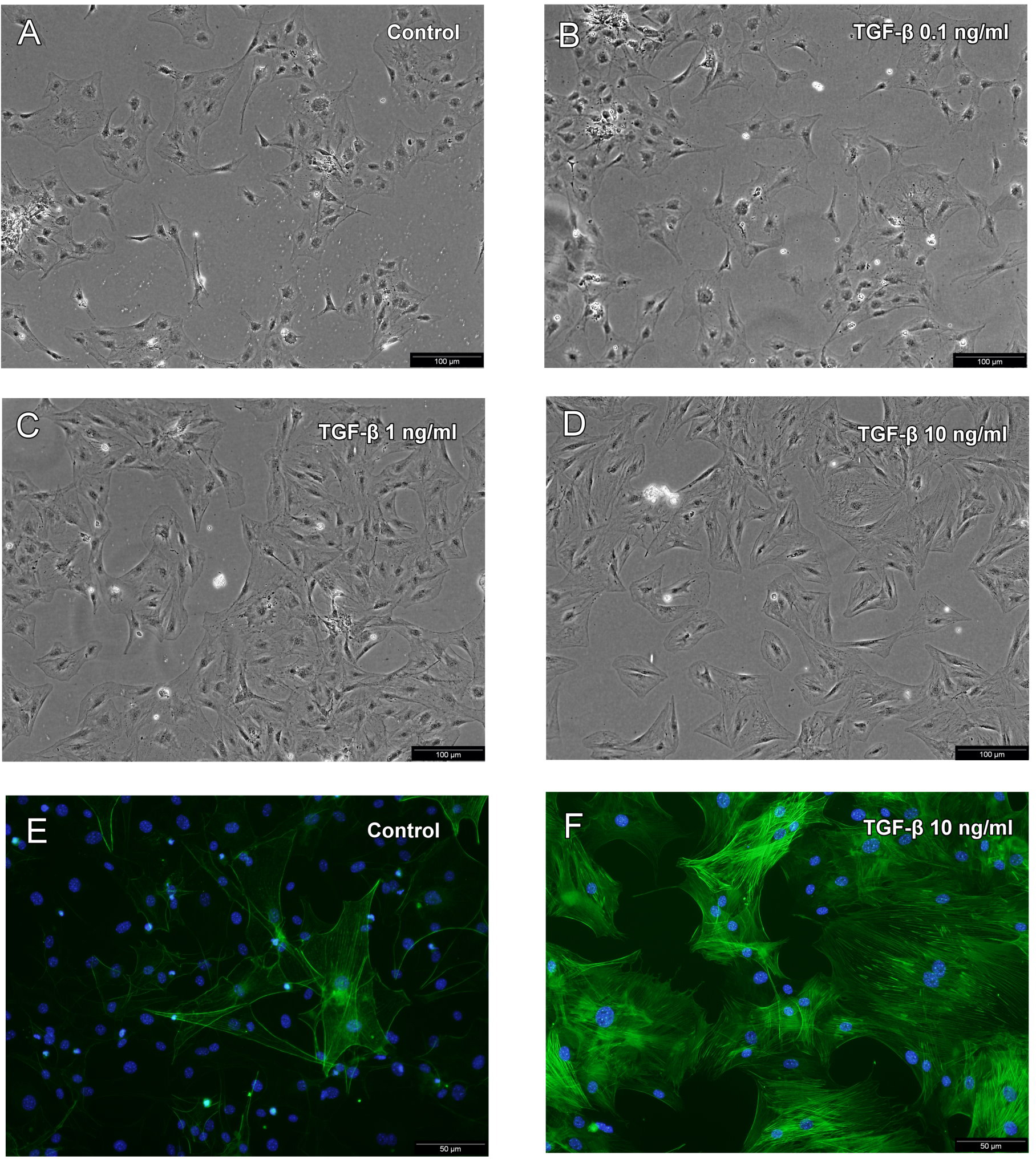
Figure 2. The changes in SSPC morphology following 24 h of treatment with different concentrations of TGF-β. A: Without TGF-β treatment, some SSPCs had the characteristics of thin spindle shapes and some showed a widened phenotype.
The cytoskeletal filaments of cells were not obvious (original magnification, ×200). B: Following treatment with 0.1 ng/ml of TGF-β, SSPCs did not change in comparison to no TGF-β treatment. C, D: SSPCs treated with 1 and 10 ng/ml of TGF-β all became broad and mostly rhombus- or triangle-shaped with prominent cytoskeletal
filaments. E, F: The expression of the α-SMA protein was determined by immunofluorescence microscopy. Nuclei were stained with DAPI (original
magnification, ×400). Representative of three independent experiments.

 Figure 2 of
Wu, Mol Vis 2015; 21:138-147.
  Figure 2 of
Wu, Mol Vis 2015; 21:138-147.