Figure 1. Slit-lamp photomicrographs of two individuals with Meesmann corneal dystrophy. A: Proband 1, left eye. Direct illumination demonstrates subtle, fine round epithelial opacities best visualized at the pupillary
border. B: Proband 1, left eye. Fluorescein staining reveals generalized stippling of the epithelium, produced by diffuse epithelial
microcysts (inset). C: Proband 2, right eye. Gray-white corneal epithelial opacities are observed in the central cornea (arrowhead), and gray-white
parallel epithelial lines are present in the inferior corneal periphery (arrow). D: Proband 2, left eye. Fluorescein staining of the peripheral, linear corneal epithelial opacities is seen.
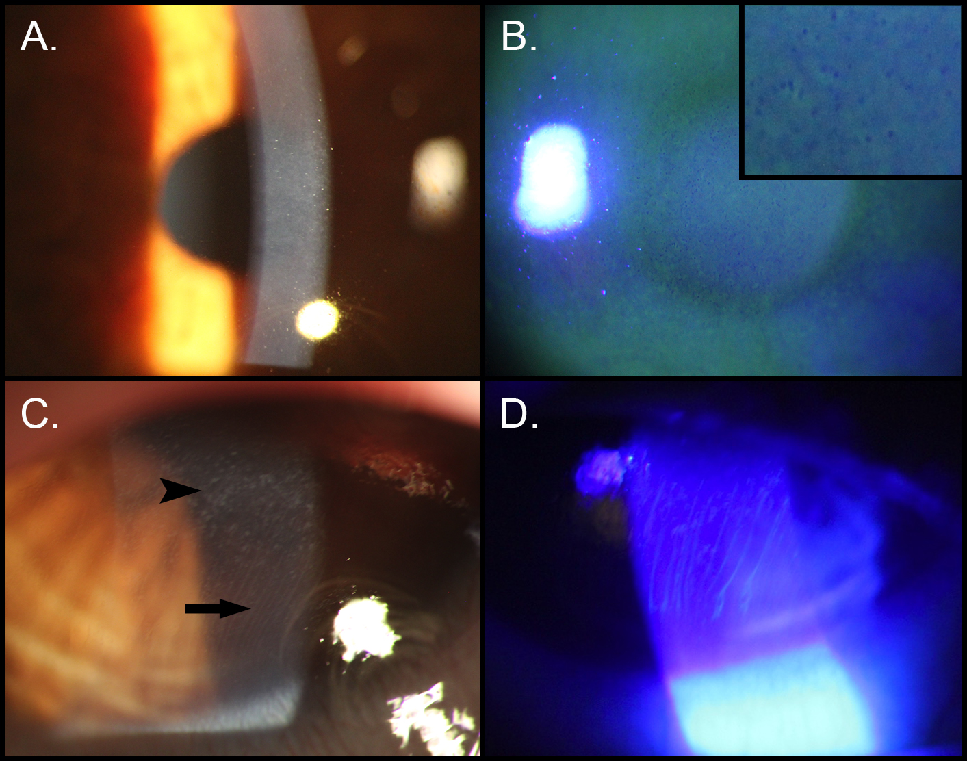
 Figure 1 of
Chen, Mol Vis 2015; 21:1378-1386.
Figure 1 of
Chen, Mol Vis 2015; 21:1378-1386.