Figure 3. Expression of ΔNp63α and PAX6. Both markers are mainly localized in the nuclei of limbal epithelial cells (LECs) cultured
on fibronectin, collagen type IV, and laminin (FCL)-coated slides. The staining is reduced in diabetic (DM) LECs compared
to the healthy (NL) LECs. Images in A and B, or in E and F, were obtained using the same exposure time. C, D, same pictures as in A and B, and G, H are the same as E and F, respectively, but with 4',6-diamidino-2-phenylindole (DAPI) nuclear counterstain. Bars=20 μm.
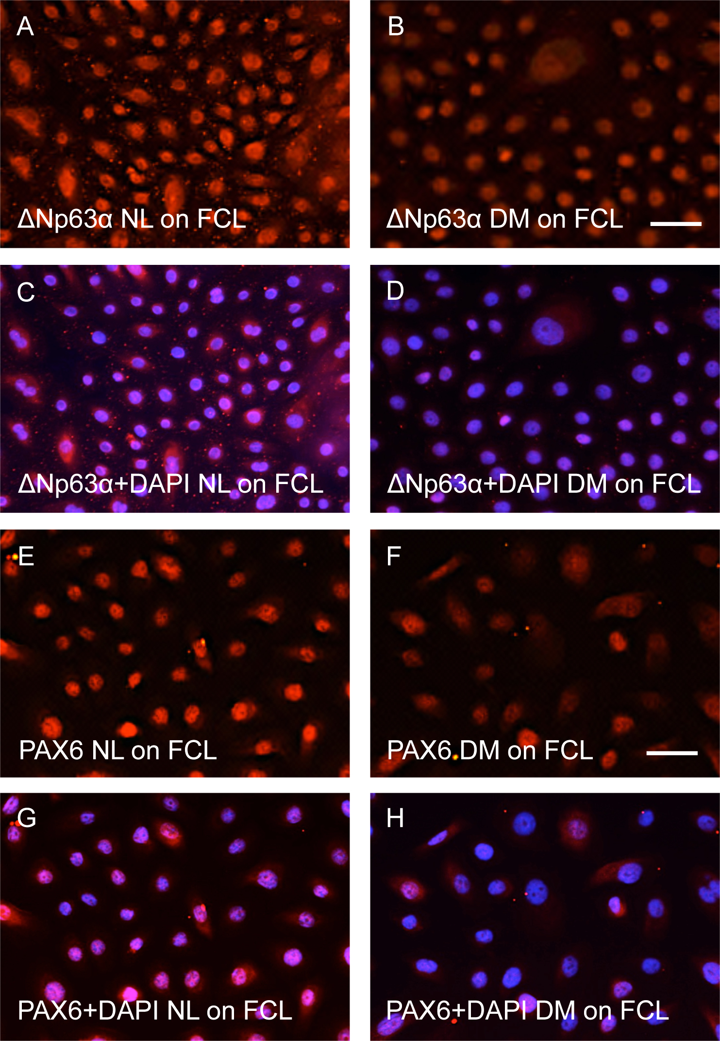
 Figure 3 of
Kramerov, Mol Vis 2015; 21:1357-1367.
Figure 3 of
Kramerov, Mol Vis 2015; 21:1357-1367.