Figure 7. Examples of applications of immunostaining on the flatmounted cornea for the study of the corneal epithelium. A, B: High magnification (40X objective) shows the characteristic cell shape of the superficial epithelial cells stained for cytokeratin
K3/12 and of the basal epithelial cells stained for CD44. C–E: Distribution of Langerhans cells, stained for S100B, in the epithelial layer. Low magnification (10X objective) easily reveals
different cell locations, most next to the limbus (dash line; D) and a few clusters migrating toward the center (E). Characteristic cell morphology is perfectly outlined using higher magnification (40X objective). For all five images, the
immunostaining is pseudocolored in green, and the nuclei, labeled by Hoechst 33,342, are pseudocolored in red.
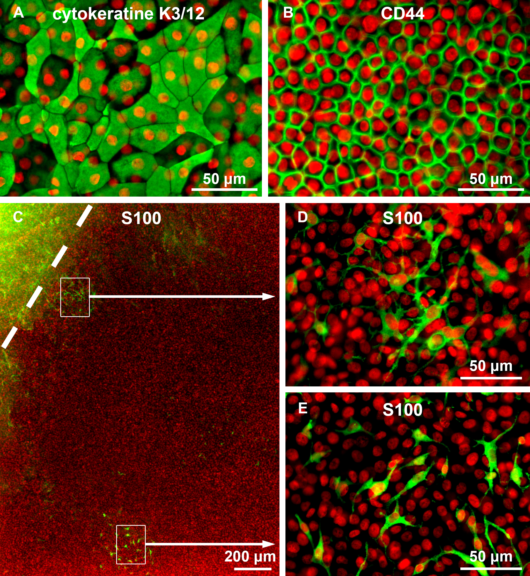
 Figure 7 of
Forest, Mol Vis 2015; 21:1345-1356.
Figure 7 of
Forest, Mol Vis 2015; 21:1345-1356.