Figure 6. Expression of neural cell adhesion molecule (NCAM) in endothelial cells (ECs) of a fresh normal cornea. A, B: Low magnification (10X objective) shows homogeneous expression of NCAMs in all ECs while medium magnification (40X objective)
reveals irregular organization of the lateral membrane where the NCAMs are located. The NCAMs are pseudocolored in green.
The nuclei, labeled by Hoechst 33342, are pseudocolored in red. C–F: High magnification (100X objective) shows continuous expression of the NCAMs at increasing depths from the apical pole of
the ECs, unveiling the increasingly irregular organization of the lateral membranes from the apical to basal pole.
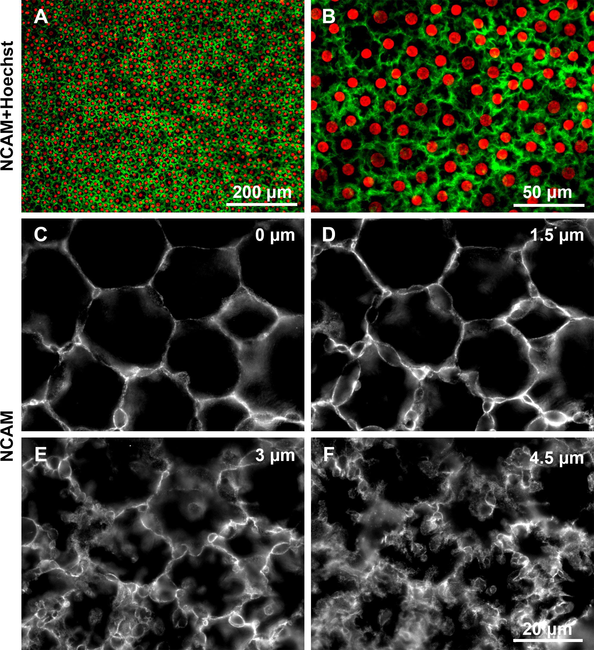
 Figure 6 of
Forest, Mol Vis 2015; 21:1345-1356.
Figure 6 of
Forest, Mol Vis 2015; 21:1345-1356.