Figure 5. Detection of the expression of the proliferation marker Ki67 in endothelial cells of corneal buttons with Fuchs endothelial
corneal dystrophy. A, B: Low magnification (10X objective) shows a few Ki67 positive cells scattered at random. Note that the nuclei morphology and
distribution of residual endothelial cells are typical of Fuchs’ corneal endothelial dystrophy (FECD), with elongated nuclei,
areas deprived of cells, and the rosette formation around Descemet excrescences forming the guttae. C, D: At high magnification (100X objective), the localization of Ki67 within the nucleolus indicates the S phase of the cell
cycle. Ki67 labeling is pseudocolored in green; the nuclei, labeled by Hoechst 33342, are pseudocolored in red.
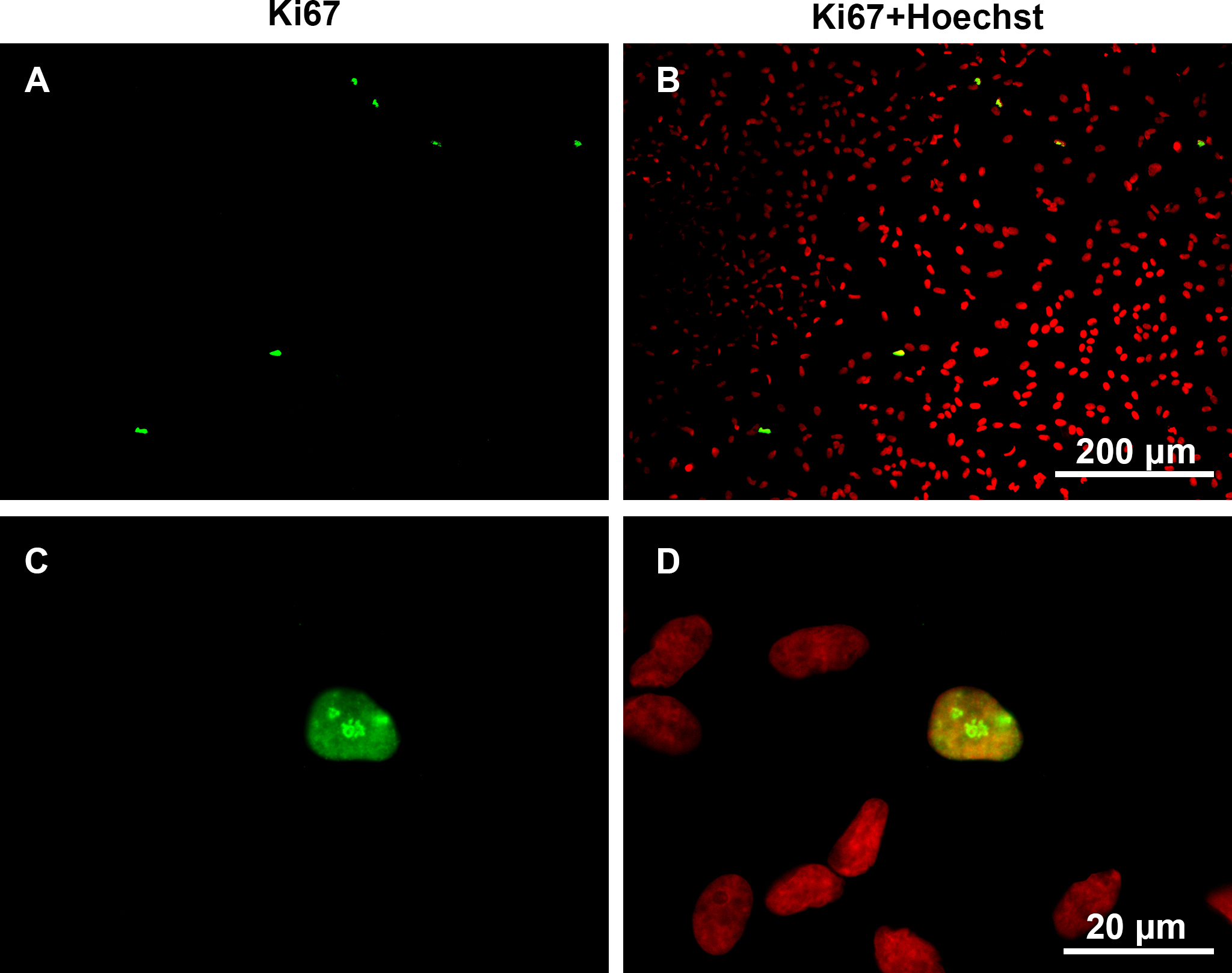
 Figure 5 of
Forest, Mol Vis 2015; 21:1345-1356.
Figure 5 of
Forest, Mol Vis 2015; 21:1345-1356.