Figure 2. Best results obtained after optimization of fixative temperature for detection in corneal ECs of flatmounted human corneas
of ZO-1 (methanol at −20 °C), hnRNP L (paraformaldehyde at room temperature [RT]), actin (methanol at 37 °C), and histone
H3 (paraformaldehyde at RT + antigen retrieval with sodium dodecyl sulfate). A: Low magnification (10X objective) shows homogeneous staining in all endothelial cells (ECs). B: High magnification (100X objective) shows the typical staining patterns for each target protein. Upper line: immunostaining
alone pseudocolored in green; lower line: merge with nuclei, labeled with Hoechst 33,342, pseudocolored in red.
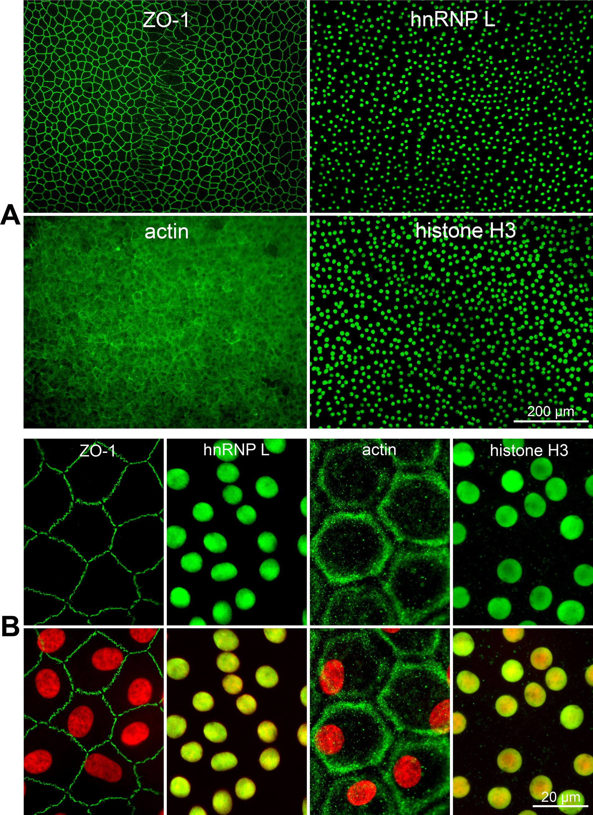
 Figure 2 of
Forest, Mol Vis 2015; 21:1345-1356.
Figure 2 of
Forest, Mol Vis 2015; 21:1345-1356.