Figure 7. Light microscopy of corneal limbus from human eye. (A) phase microscopy image. (B) fluorescence microscopy image of same section. Positive immunofluorescence was detected in the sub-epithelial stroma in
the region of stromal cell–epithelial cell interactions only with the CS/DS motif-specific antibody 6C3. The scale bar represents
50 µm.
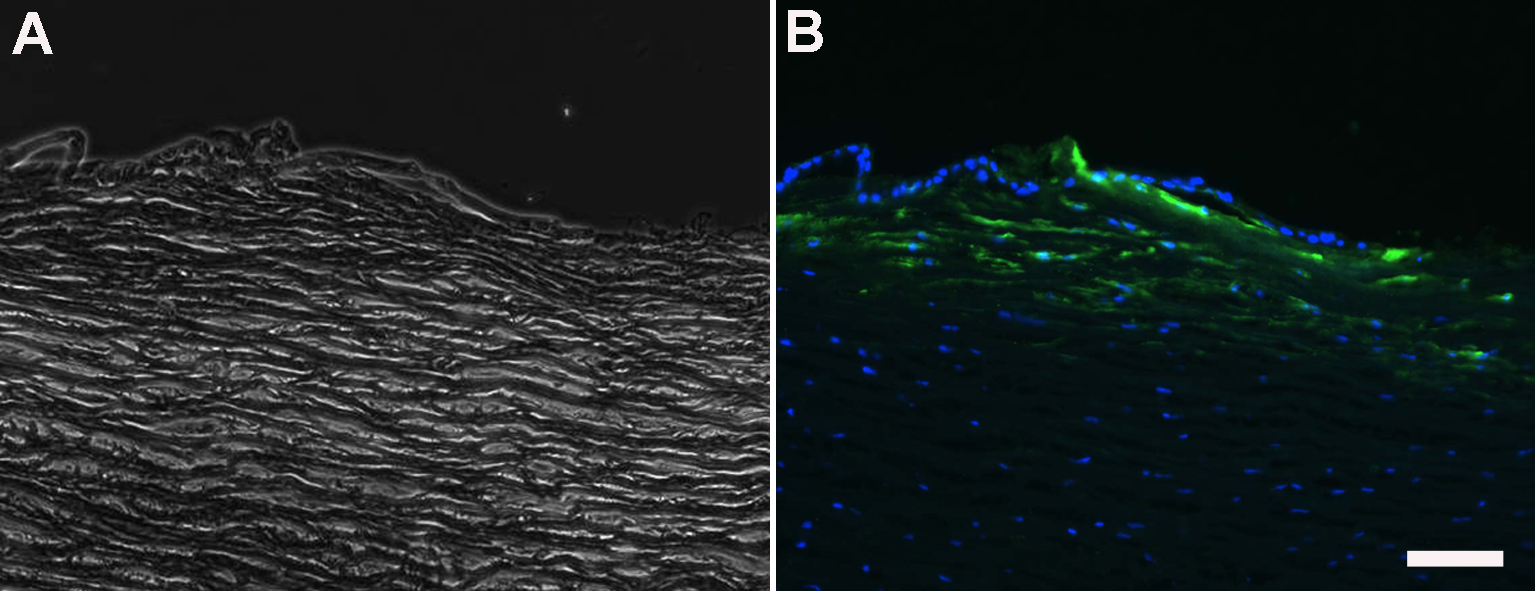
 Figure 7 of
Yamada, Mol Vis 2015; 21:1328-1339.
Figure 7 of
Yamada, Mol Vis 2015; 21:1328-1339.