Figure 6. Immunolocalization of chondroitin sulfate proteoglycan in the rabbit corneal limbus using five sulfation motif-specific monoclonal
antibodies: 3B3, 3C5, 4C3, 6C3, and 7D4. Phase contrast images are shown in the left-hand panels (A, D, G, J, M, and P); the center panels (B, E, H, K, N, and Q) show native epitope localization without section pretreatment. Antibody 6C3 labels the matrix subjacent to the limbal epithelial
basement membrane (K, arrows). The right-hand panels (C, F, I, L, O, and R) show validation of the results by removal of native epitopes by section pretreatment with the chondroitinase ABC enzyme,
and thus, the loss of the 6C3 signal (L). In some cases, neoepitopes are generated by enzyme treatment, as with antibody 3B3. The central cornea is toward the left
in all panels, but toward the right in the controls (P, Q, and R). The scale bar represents 100 µm.
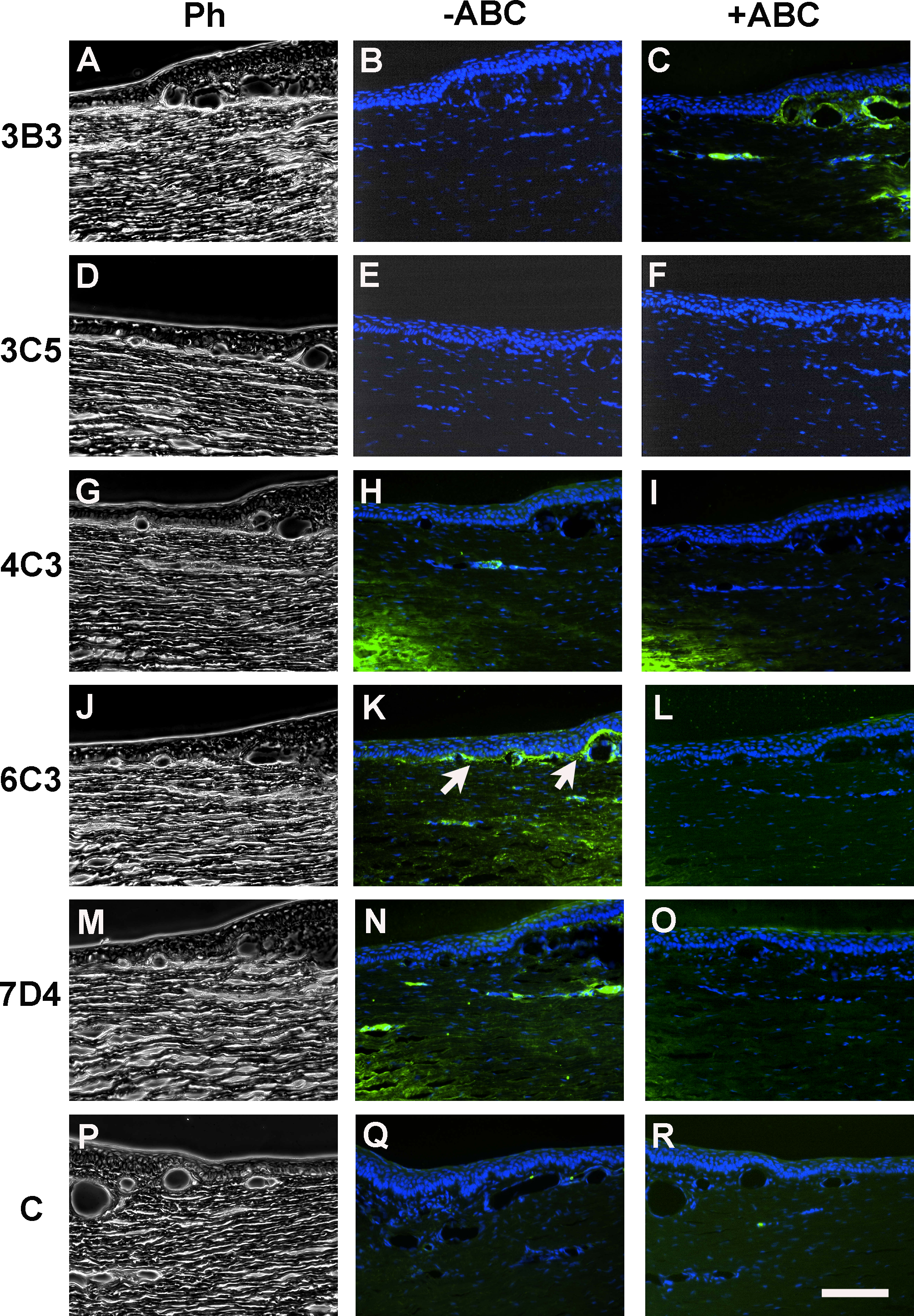
 Figure 6 of
Yamada, Mol Vis 2015; 21:1328-1339.
Figure 6 of
Yamada, Mol Vis 2015; 21:1328-1339.