Figure 5. Three-dimensional reconstruction of the rabbit corneal limbal basement membrane zone using automated and manual segmentation
techniques with Amira 5.6 software. Mesenchymal cells colored in blue and purple make associations with basal epithelial cells,
in green. A superficial stromal capillary (orange) can also be seen. The scale bar represents 4 µm.
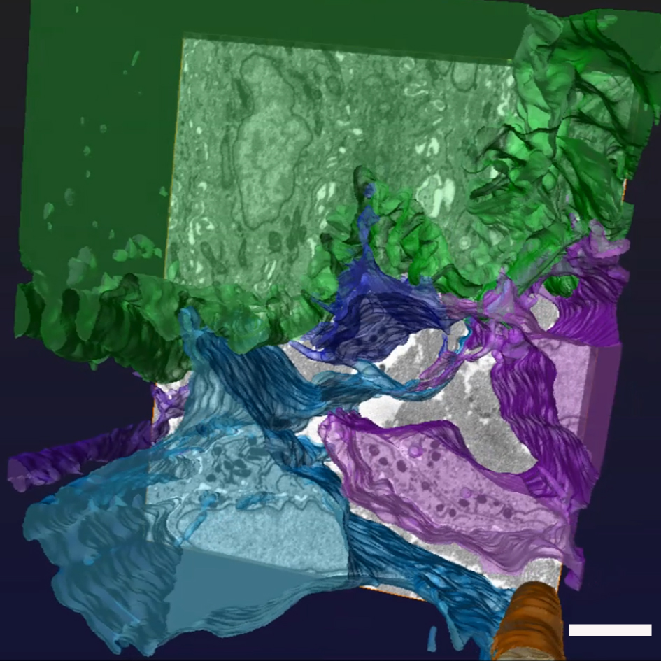
 Figure 5 of
Yamada, Mol Vis 2015; 21:1328-1339.
Figure 5 of
Yamada, Mol Vis 2015; 21:1328-1339.