Figure 4. Three-dimensional reconstructions in ImageJ 3D Viewer of the epithelial basement membrane zone of the rabbit limbal cornea
from serial block face scanning electron microscopy. A: A blood vessel can be seen in the superficial stroma (asterisk), below mesenchymal cells that extend numerous cytoplasmic
processes (arrows) distally to contact the basal epithelial cells. The scale bar represents 4 µm. B: Mesenchymal cell processes form diffuse associations with epithelial cells, occasionally appearing to extend between adjacent
cells (arrow). The scale bar represents 4 µm.
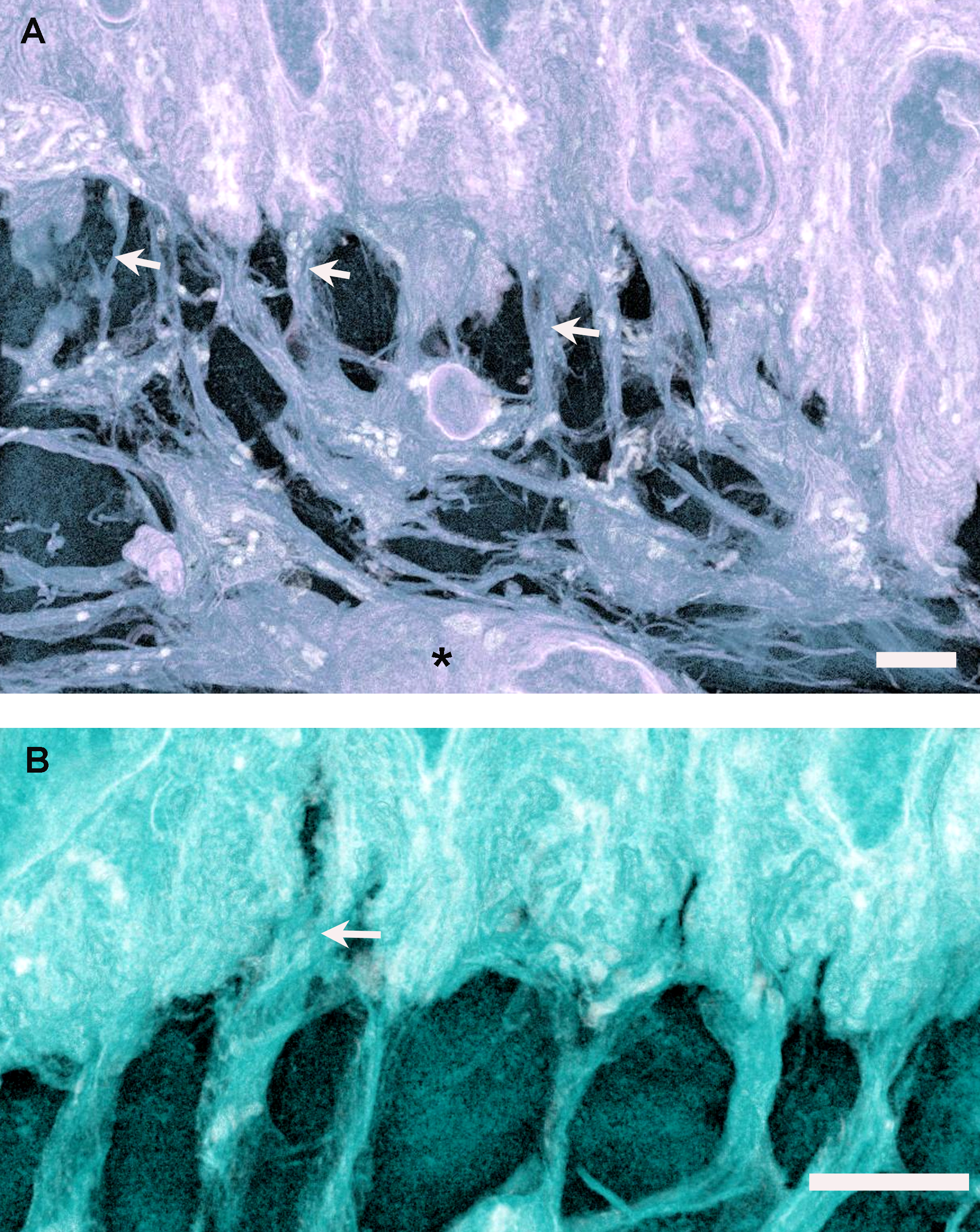
 Figure 4 of
Yamada, Mol Vis 2015; 21:1328-1339.
Figure 4 of
Yamada, Mol Vis 2015; 21:1328-1339.