Figure 3. Reversed-contrast backscatter electron images from serial block face scanning electron microscopy of the limbal epithelial
basement membrane zone in the rabbit cornea. A–D show mesenchymal–epithelial cell interactions at 20 image intervals, in approximately 1 µm increments. A: A subepithelial mesenchymal cell extends a process toward the basement membrane (arrow). B–D: Over the subsequent three microns, the process extends deeply into the recess formed by the lobed basal epithelial cells
(arrows), appearing to make direct contact through the basal lamina (D). The scale bar represents 5 µm.
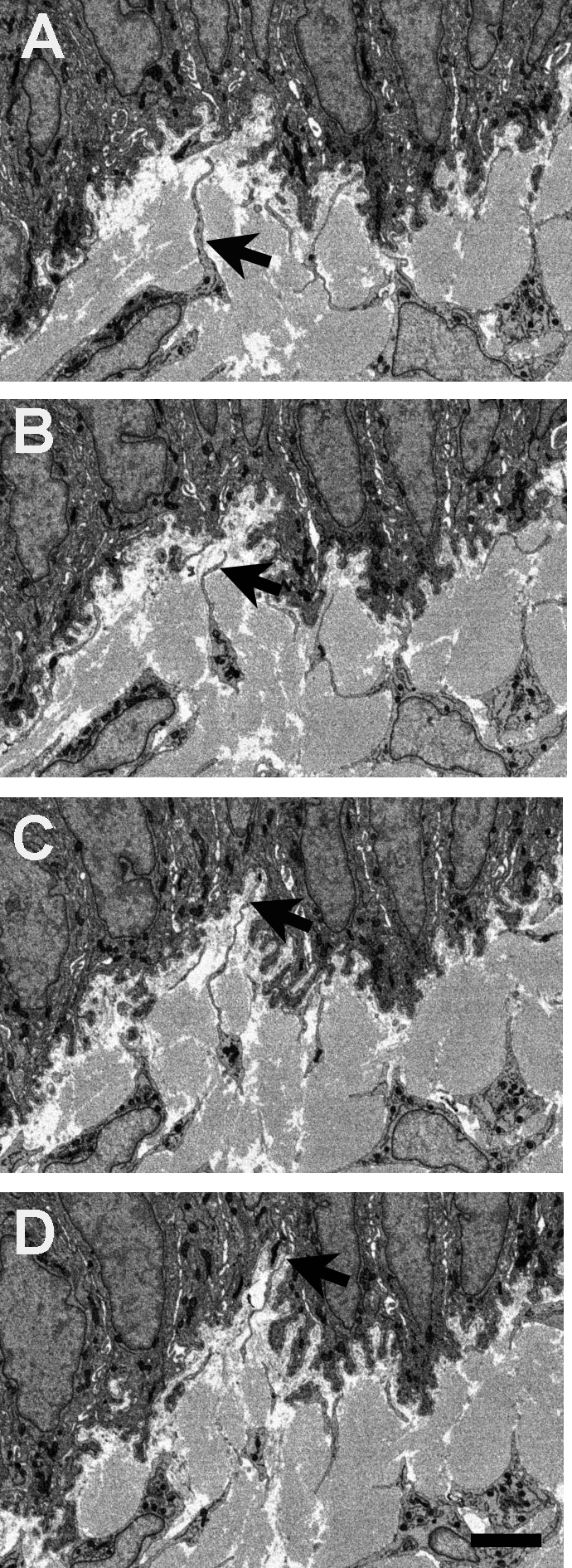
 Figure 3 of
Yamada, Mol Vis 2015; 21:1328-1339.
Figure 3 of
Yamada, Mol Vis 2015; 21:1328-1339.