Figure 2. Transmission electron microscopy of the epithelial basement membrane zone in the rabbit corneal limbus. A: Basal epithelial cells surround a small rounded cell with a high nuclear to cytoplasm ratio (asterisk). Lobed processes
of basal cells, with associated basal lamina (arrow), project into the superficial stroma. Numerous cytoplasmic extensions
from the mesenchymal cells make contact with the basal lamina and insert between the basal cell processes (arrowheads). The
scale bar represents 2 µm. B: Detail of the contact between the mesenchymal cell (M) and the epithelial cell process (E), where the basal lamina appears
discontinuous (arrowhead). The scale bar represents 0.5 µm.
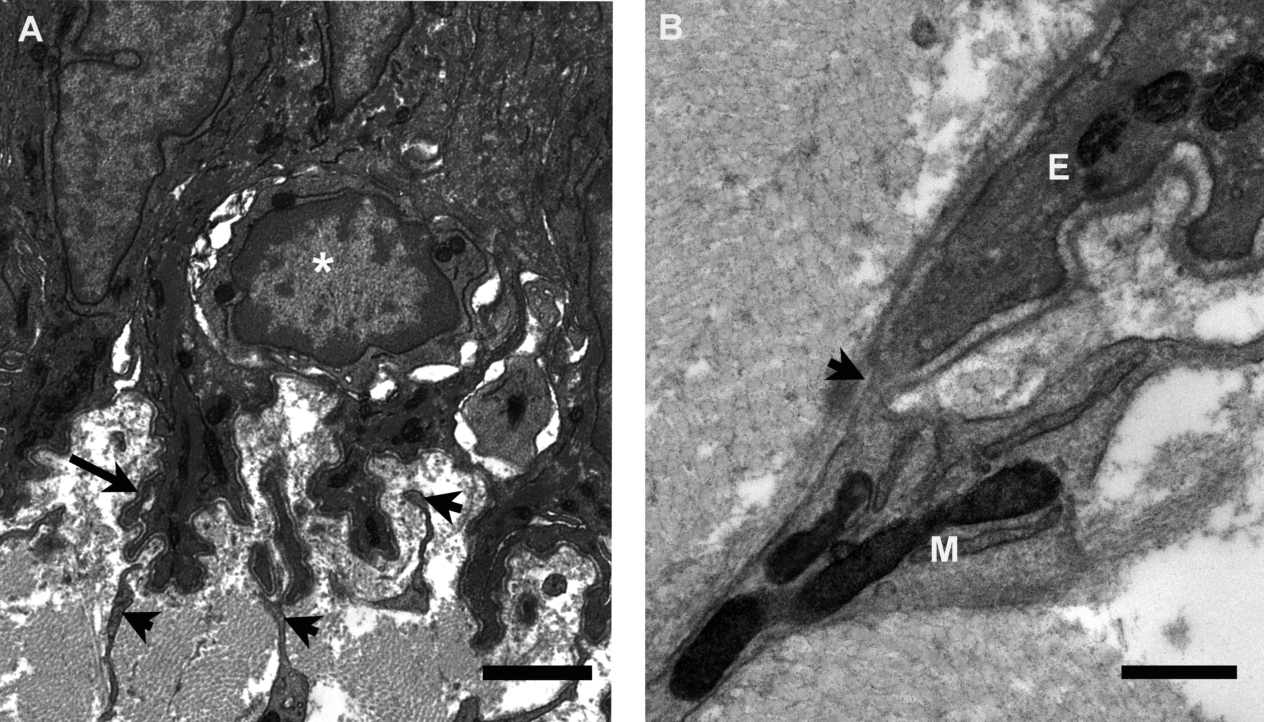
 Figure 2 of
Yamada, Mol Vis 2015; 21:1328-1339.
Figure 2 of
Yamada, Mol Vis 2015; 21:1328-1339.