Figure 1. Toluidine blue-stained semithin sections of rabbit corneal limbus. (A) Superior limbal location, (B) Nasal, (C) Inferior and (D) Temporal. Palisades of Vogt are absent although the epithelial basement membrane has an irregular profile. Subepithelial
mesenchymal cells, adjacent to capillaries (asterisks) in the superficial stroma, extend the processes distally to make contact
with the basal epithelial cells (white arrows). The irregular dark lines (black arrowheads) are artifactual microfolds in
the resin section, readily distinguished from the blue-stained cell processes. The central cornea is toward the right of the
region illustrated in A and B and toward the left in C and D. The scale bar represents 100 µm.
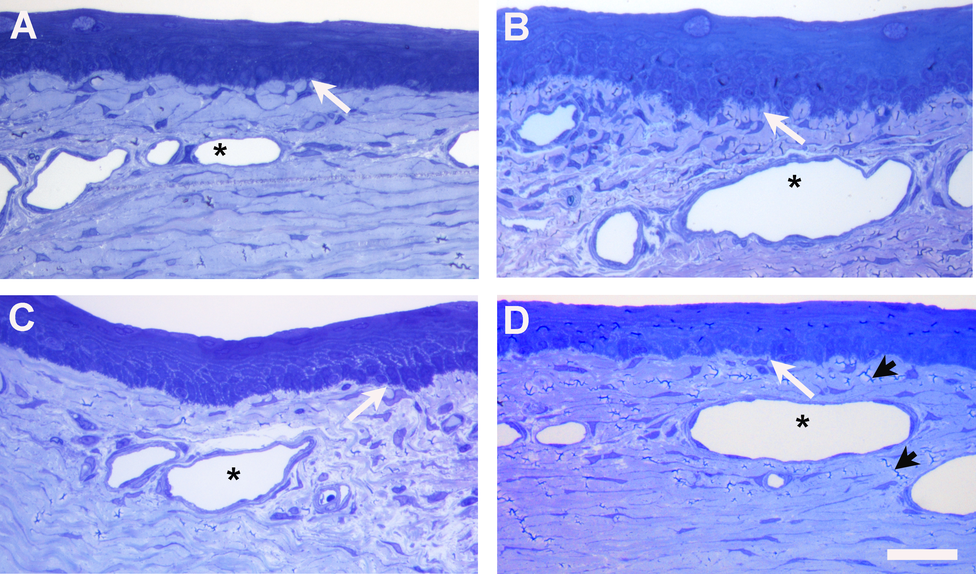
 Figure 1 of
Yamada, Mol Vis 2015; 21:1328-1339.
Figure 1 of
Yamada, Mol Vis 2015; 21:1328-1339.