Figure 4. Perlecan immunocytochemistry of corneal stromal cells cultured under different conditions. Perlecan (red) was most highly
expressed in localized intracellular structures in keratocytes whereas in the myofibroblasts perlecan was detected more diffusely
throughout the cell (white arrowheads). Fibroblasts had little, if any, detectable expression of perlecan protein. The isotypic
control immunoglobulin (IgG) did not yield staining under any of the culture conditions, as is shown for myofibroblasts. Magnification=400X.
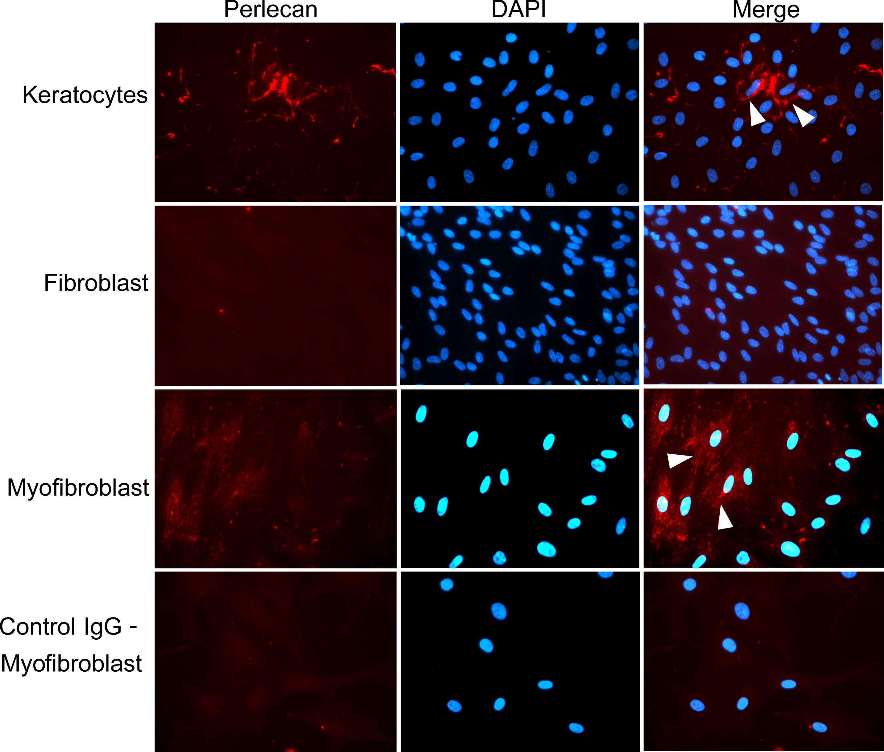
 Figure 4 of
Santhanam, Mol Vis 2015; 21:1318-1327.
Figure 4 of
Santhanam, Mol Vis 2015; 21:1318-1327.