Figure 3. Nidogen-2 immunocytochemistry of corneal stromal cells under different culture conditions. The expression of nidogen-2 protein
(green) is seen to be highest in the perinuclear organelle, whereas in myofibroblasts nidogen-2 protein expression appears
as a meshwork-like pattern throughout the cell body (white arrowheads). Fibroblasts have little, if any, nidogen-2 protein
expression, except the perinuclear organelle staining, as was observed in keratocytes. Isotypic control immunoglobulin (IgG)
staining was negative under all cell conditions, as shown for the myofibroblasts. Magnification=400X.
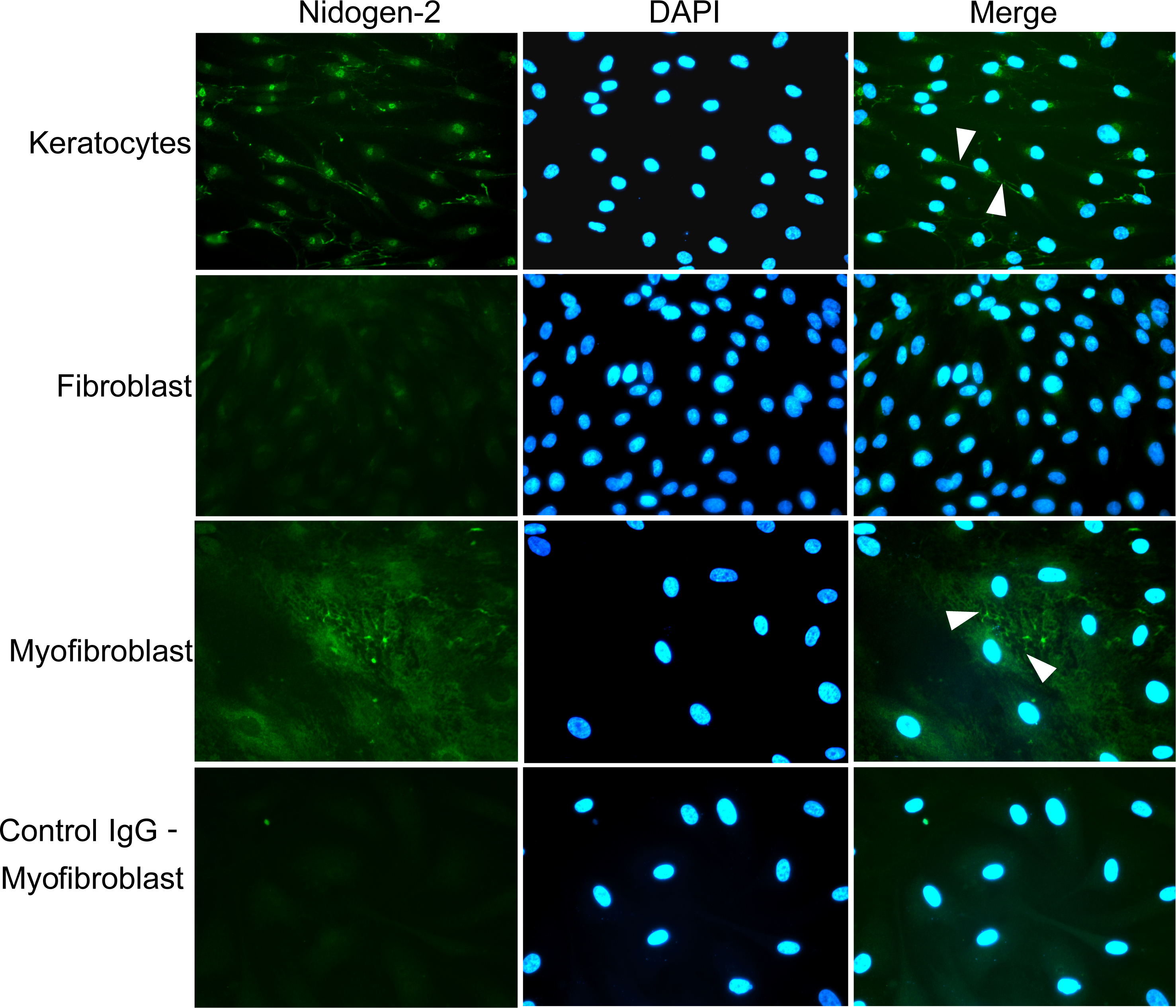
 Figure 3 of
Santhanam, Mol Vis 2015; 21:1318-1327.
Figure 3 of
Santhanam, Mol Vis 2015; 21:1318-1327.