Figure 2. Broad inflammation and neovascularization characterize affected corneas. Central corneas from affected HLA-B27/DKO animals
stain positive for a panel of markers, including CD31 (A, B), CD4 (C, D), CD8 (E, F), F4/80 (G, H), and Ly-6G (I, J), as compared to negative WT controls (n = 6). (Bar indicates 50 μM).
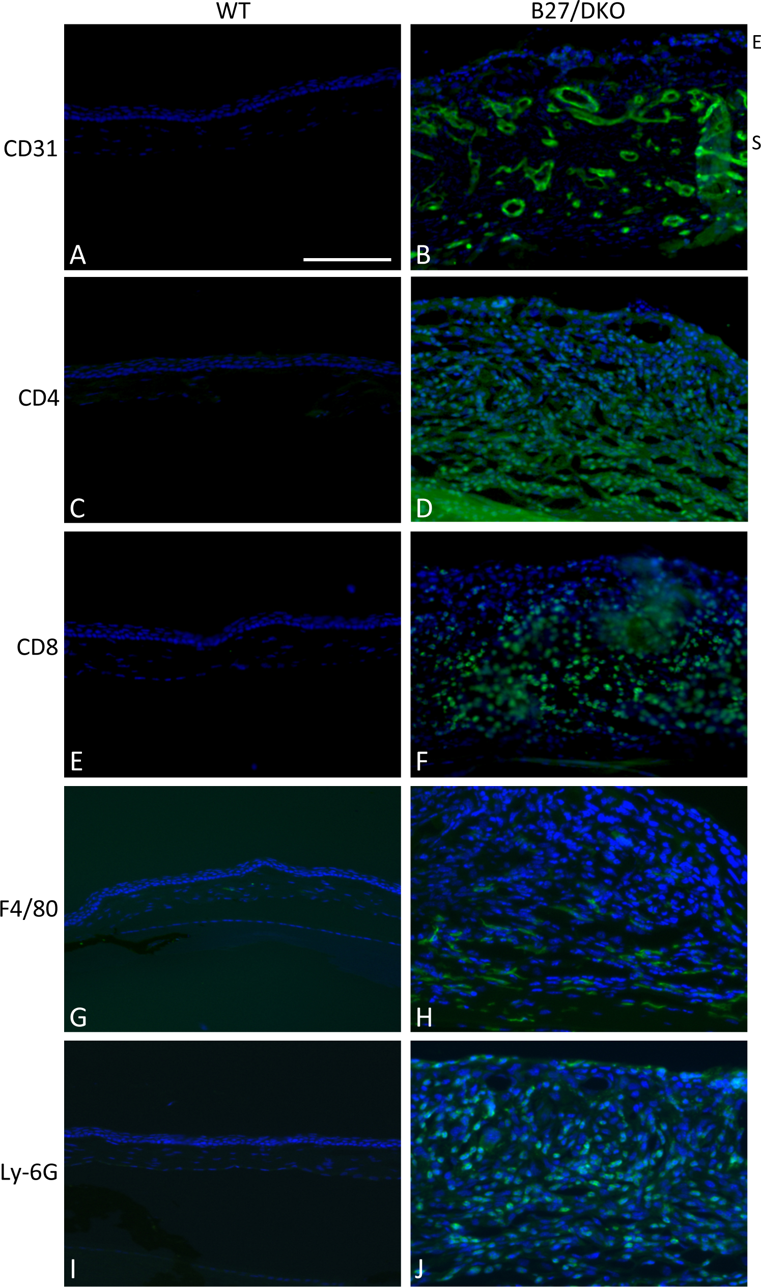
 Figure 2 of
Lin, Mol Vis 2015; 21:131-137.
Figure 2 of
Lin, Mol Vis 2015; 21:131-137.