Figure 1. Rare corneal opacities observed in HLA-B27/DKO mice. A: A clear eye from an HLA-B27/DKO mouse. B: The contralateral eye developed a severe central corneal opacity. C: H&E staining of the periphery of an affected cornea shows some inflammatory infiltrates but limited tissue disruption. D: H&E staining of the central cornea shows a damaged but intact epithelium and a severely disrupted stroma, accompanied by
a massive number of tissue infiltrates and neovascularization (n = 6). (E: epithelium, S: stroma, bar indicates 50 μM).
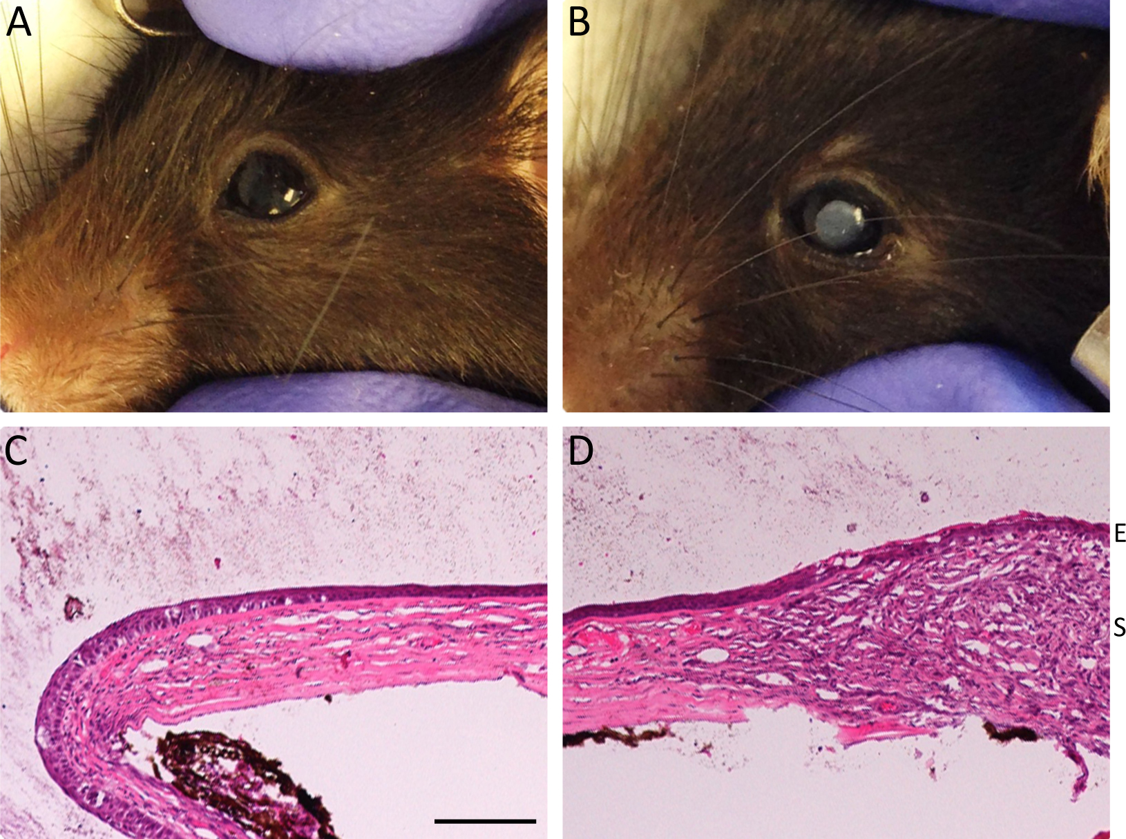
 Figure 1 of
Lin, Mol Vis 2015; 21:131-137.
Figure 1 of
Lin, Mol Vis 2015; 21:131-137.