Figure 8. Immunohistochemistry analysis of the inner retina with PSD-95, calbindin, and PKC-α. A: Primary antibodies for PSD-95 (1:500) and calbindin (1:1,000) were followed by Alexa Fluor donkey anti-mouse 488 (1:500)
and Alexa Fluor goat anti-rabbit 568 (1:500), respectively, for dual localization immunoreactive stain. The synapses (green)
between the photoreceptor and horizontal cells (red) were normal for all genotypes. B: Primary antibodies to PSD-95 (1:500) and PKC-α (1:1,000) followed by Alexa Fluor donkey anti-mouse 488 (1:500) and Alexa
Fluor goat anti-rabbit 568 (1:500), respectively, for the dual localization immunoreactive stain. The synapses (green) between
the photoreceptor and bipolar cells (red) for most retinas were normal for all genotypes. C: Occasionally, bipolar cells in the B6N background retinas appear to form ectopic synapses in the outer nuclear layer. Images
taken with a 40X objective lens at 2X zoom (scale bar, 50 µm). Magnified images taken with a 63X objective lens (scale bar,
10 µm). ONL=outer nuclear layer; OPL=outer plexiform layer; INL=inner nuclear layer.
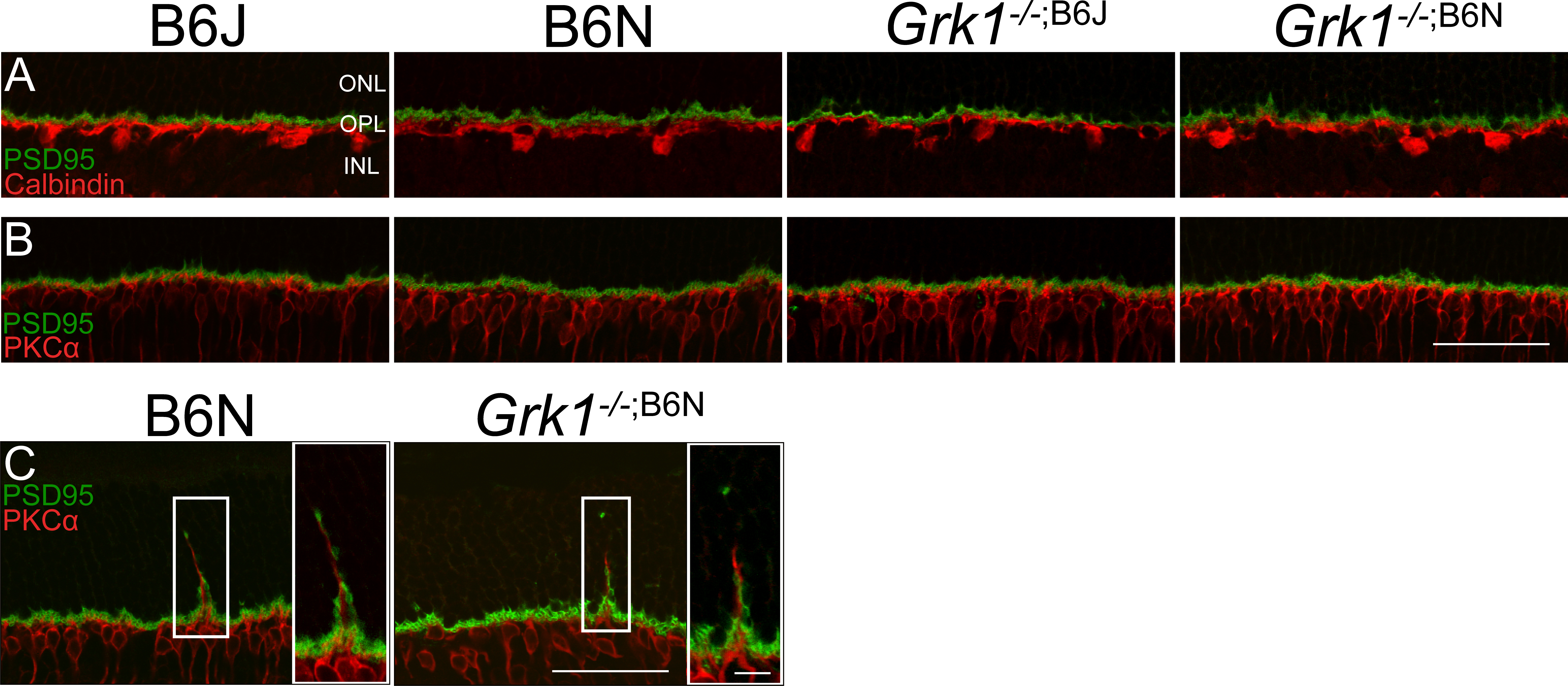
 Figure 8 of
Pak, Mol Vis 2015; 21:1281-1294.
Figure 8 of
Pak, Mol Vis 2015; 21:1281-1294.