Figure 6. Immunohistochemistry analysis of retinal ZO-1. Primary antibody to ZO-1 (1:1,000) followed by secondary antibody Alexa Fluor
donkey anti-rabbit 488 (1:500) identifies immunohistochemical staining in mouse retina sections at 1 month (A–D) and 3 months (E–H) localized to the OLM. A, C, E, G: The OLM in the B6J background retinas is unbroken. B, D, F, H: The OLM in the B6N background retinas is discontinuous. The top-right corners show higher magnification images. Lower magnification
images were taken with a 20X objective lens (scale bar, 50 µm); higher magnification images were taken at 100X objective (scale
bar, 20 µm). OLM=outer limiting membrane; ONL=outer nuclear layer.
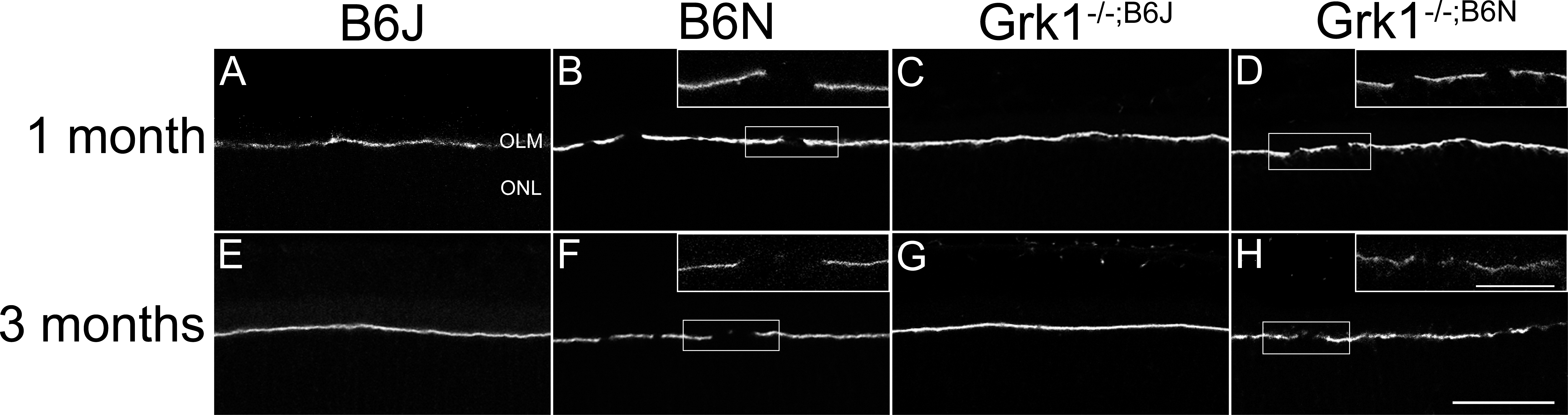
 Figure 6 of
Pak, Mol Vis 2015; 21:1281-1294.
Figure 6 of
Pak, Mol Vis 2015; 21:1281-1294.