Figure 5. Immunohistochemistry analysis of retinal GFAP. Vertical sections were immunologically stained with the primary antibody for
glial fibrillary acidic protein (GFAP) (1:1,000) followed with the secondary antibody Alexa Fluor donkey anti-rabbit 488 (1:500)
for 1-month-old (A–D) and 3-month-old (E–H) retinas. A and E: Immunoreactive GFAP is restricted to the innermost layer of the retina in the B6J retinas. B and F: GFAP is predominantly restricted to the nerve fiber layer (NFL) but with occasional fibrils extending into the IPL (C and G). GFAP is moderately upregulated in the processes extended into the INL and some into the ONL. D and H: Widespread GFAP upregulation in the processes extending into the ONL. Images were taken using a 20X objective lens (scale
bar, 50 µm). ONL=outer nuclear layer; OPL=outer plexiform layer; INL=inner nuclear layer; IPL=inner plexiform layer; GCL=ganglion
cell layer.
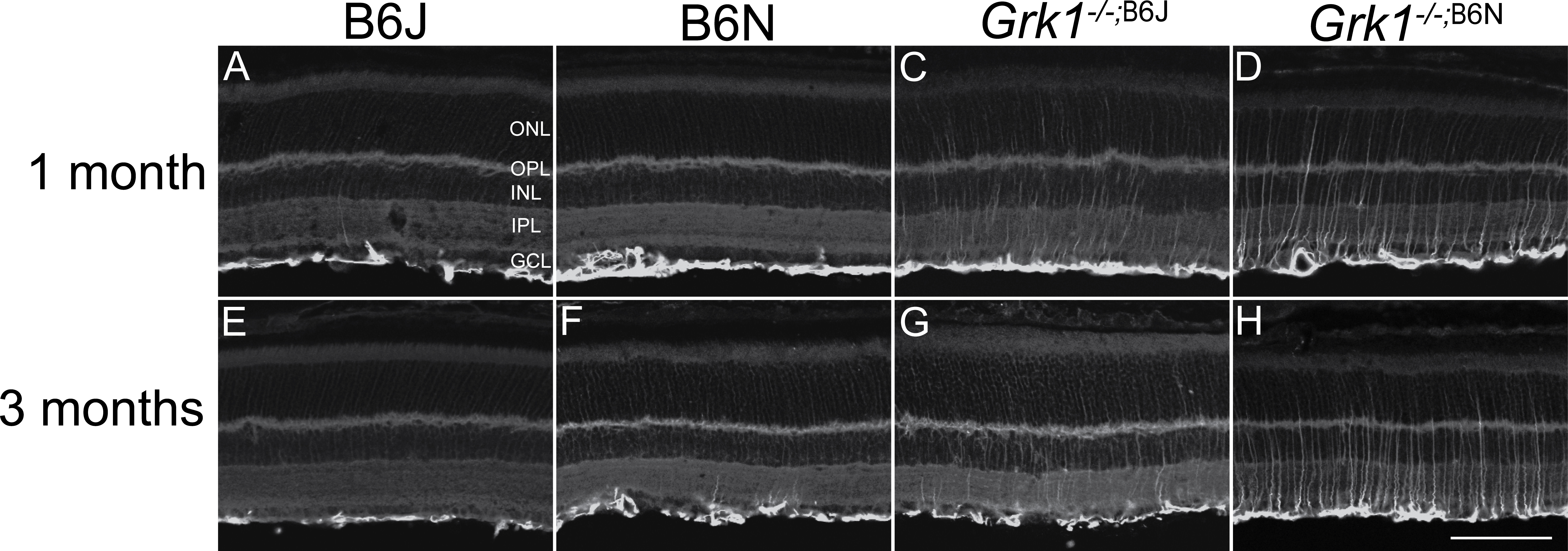
 Figure 5 of
Pak, Mol Vis 2015; 21:1281-1294.
Figure 5 of
Pak, Mol Vis 2015; 21:1281-1294.