Figure 4. Superior and inferior retina TUNEL analysis. A and B are representative plots of terminal deoxynucleotidyl transferase-mediated dUTP nick-end labeling (TUNEL)-positive nuclei
at defined points in the retina at (A) 1 and (B) 3 months. Mice were exposed to 24 h of 1,000 lux light and were immediately euthanized. TUNEL staining was performed on
10 µm thick sections, and counts were taken from a 0.01 mm2 area every 0.4 µm from the OD. The Grk1−/−;B6J (black asterisk) and Grk1−/−;B6N (red asterisk) retinas have significantly increased levels of TUNEL-positive nuclei compared to the B6J retinas. The purple
asterisk shows Grk1−/−;B6N significance compared to that of B6N. The level of significance of the Grk1−/−;B6J retinas compared to the B6N retinas is identical in comparison to the B6J retinas (A). Grk1−/−;B6N TUNEL is significantly increased against Grk1−/−;B6J at one point in the inferior retina (A, −0.4 mm, green asterisk). C and D are total TUNEL-positive counts in the inferior and superior retina at (C) 1 and (D) 3 months. The Grk1−/−;B6J and Grk1−/−;B6N retinas are significantly increased compared to the B6J and B6N retinas (*p<0.05, **p<0.01, ***p<0.001, ****p<0.0001). OD=optic
disc.
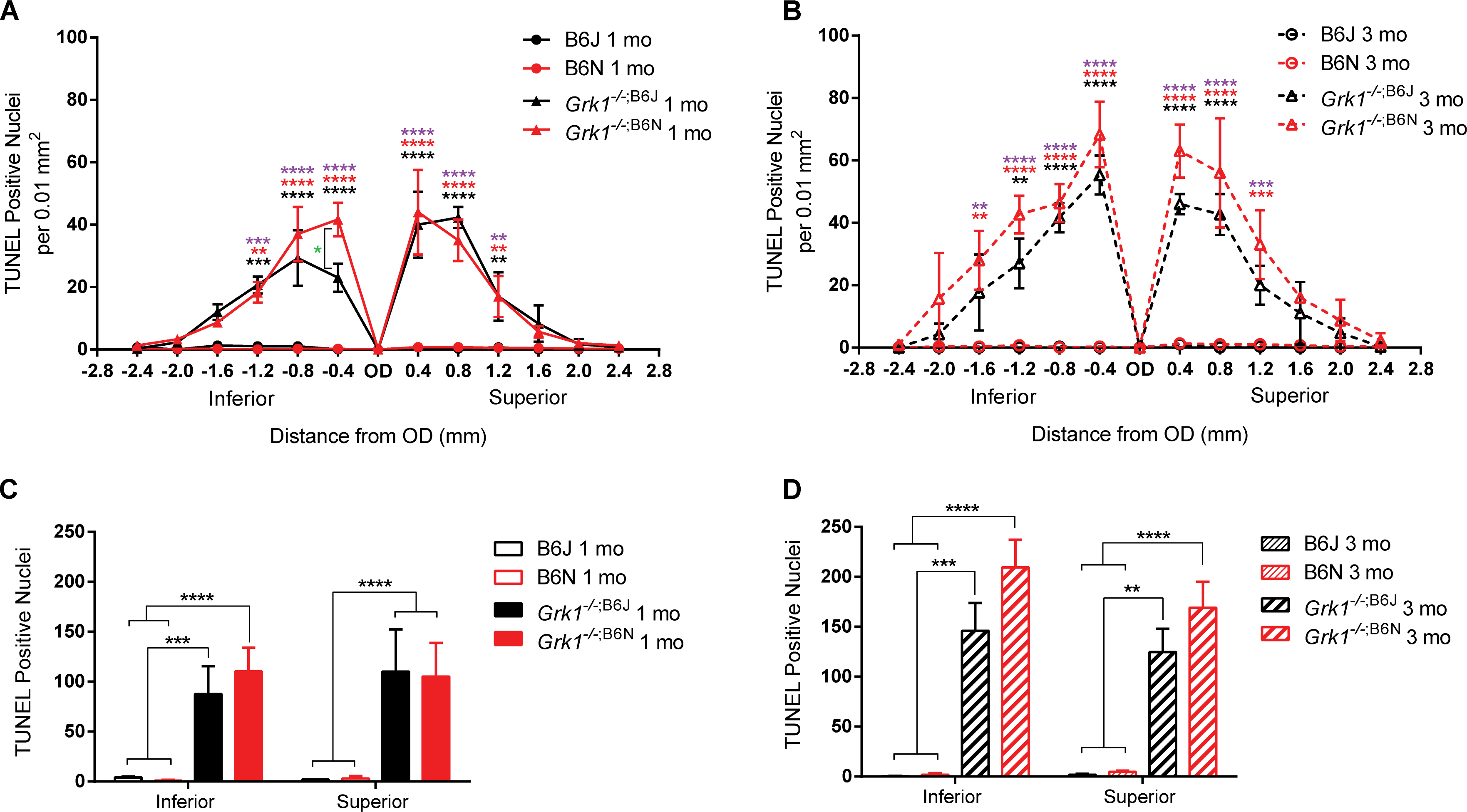
 Figure 4 of
Pak, Mol Vis 2015; 21:1281-1294.
Figure 4 of
Pak, Mol Vis 2015; 21:1281-1294.