Figure 2. Microanatomy and outer nuclear layer thickness. Retinal nuclei of the ONL stained with TO-PRO-3 and imaged for 1-month-old
(A–D) and 3-month-old (E–H) animals. The B6J and Grk1−/−;B6J retinas have well-organized outer nuclear layers while the B6N (B and F) and Grk1−/−;B6N (D and H) retinas show disorganization in the outer portion of the ONL, with holes forming at the outer edge of the ONL (arrowheads;
scale bar, 50 µm). ONL=outer nuclear layer; OPL=outer plexiform layer; INL=inner nuclear layer.
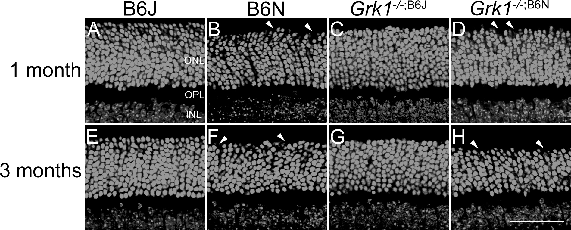
 Figure 2 of
Pak, Mol Vis 2015; 21:1281-1294.
Figure 2 of
Pak, Mol Vis 2015; 21:1281-1294.