Figure 1. Representative images of histopathology and TEM in FECD. A: Photomicrograph of hematoxylin and eosin staining (20X) reveals stromal edema with thickened Descemet’s and retrocorneal
membrane. B: Descemet’s membrane thickening of the DSAEK specimen (40X). C: Photomicrograph of hematoxylin and eosin staining (20X) of a control. D: Transmission electron microscopy (TEM) showing Descemet’s membrane of typical Fuchs endothelial corneal dystrophy (FECD).
Four regions are distinguished: anterior banded, non-banded, posterior banded, and the fibrillar region (scale bar, 2 µm).
E: Enlarged view of the posterior stroma with guttae-like deposits. F: Enlarged view of the stroma showing degenerative changes and guttae-like excrescences of spindle-shaped wide-space collagen
(scale bar, 200 nm).
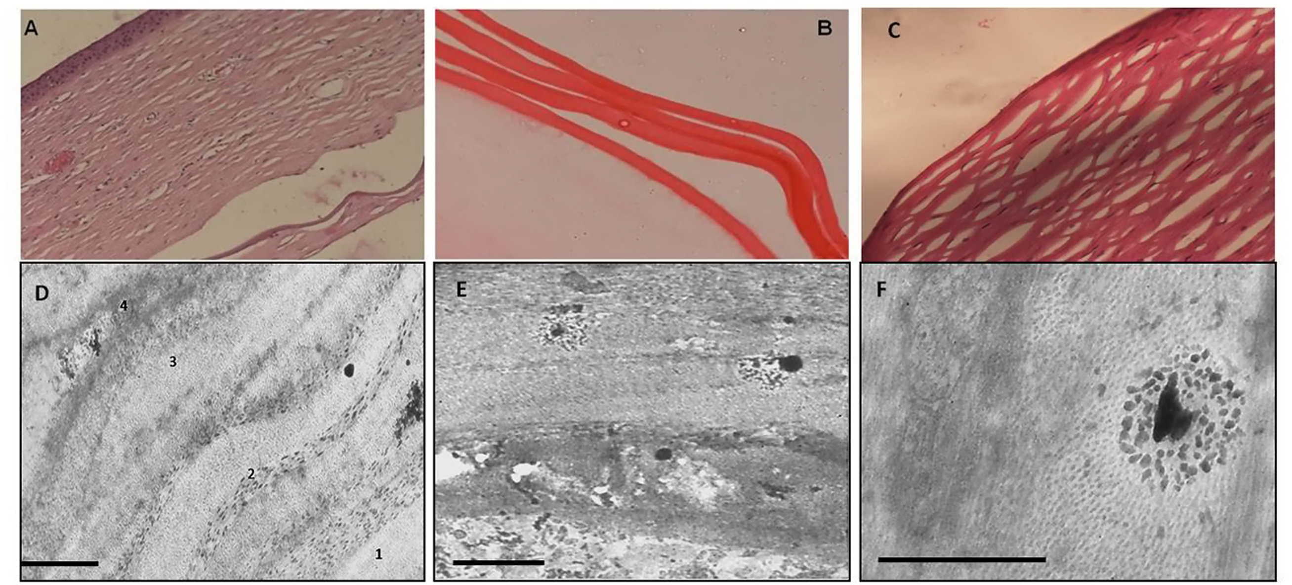
 Figure 1 of
Gupta, Mol Vis 2015; 21:1252-1260.
Figure 1 of
Gupta, Mol Vis 2015; 21:1252-1260.