Figure 3. Effect of CDECM on the detachment of corneal epithelial cells. Stained corneas from the experimental NOD.B10.H2b mice. A: Hematoxylin and eosin (H&E) staining: The corneas of the NOD.B10.H2b mice were stained 10 days (10D) after the instillation of PBS or chondrocyte-derived extracellular matrix (CDECM) five times
per day for 10D. The arrows indicate the detached corneal epithelial cells. Scale bar=100 μm. B: The quantitative data are presented as mean ± standard deviation (SD). *p<0.05 versus the value in the control. #p<0.05 versus the corresponding value in the DS 10D group. DS 10D, desiccation stress for 10 days; PBS 10D, PBS group after
10 days of instillation; CDECM 10D, CDECM group after 10 days of instillation.
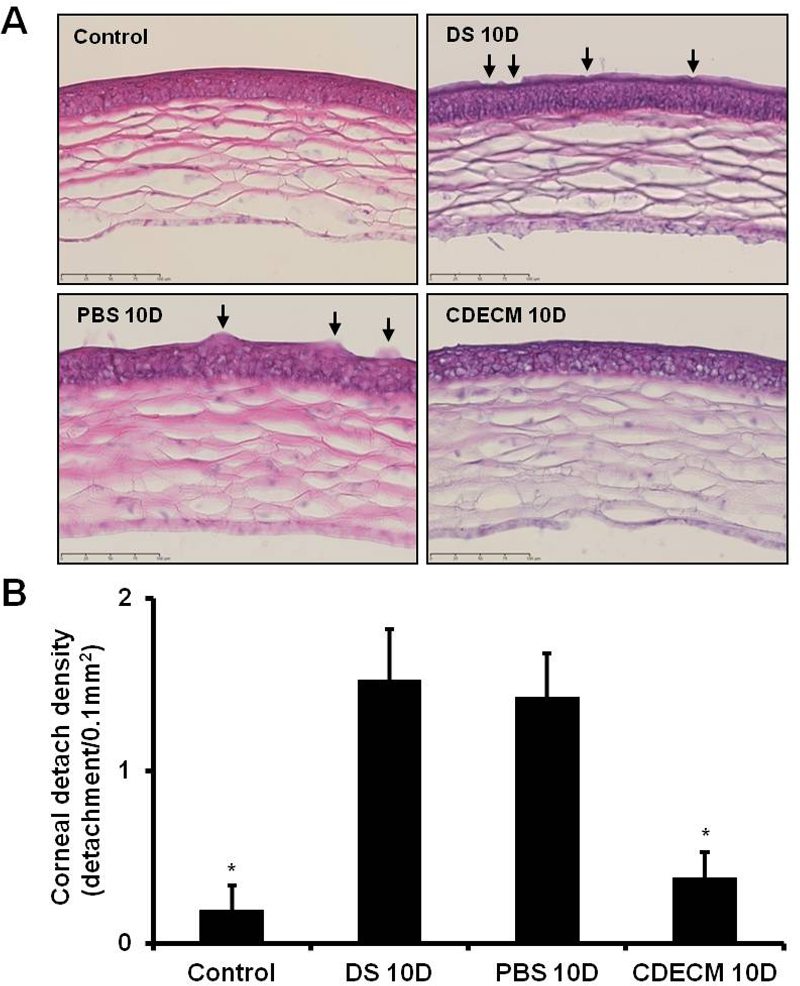
 Figure 3 of
Kim, Mol Vis 2015; 21:1210-1223.
Figure 3 of
Kim, Mol Vis 2015; 21:1210-1223.