Figure 3. Histopathological findings in a 15-year-old female patient (patient 6). A: Hematoxylin and eosin stain. B: Alcian blue stain. C: Periodic acid-Schiff stain. D: Congo red stain. E: Masson’s trichrome stain. Basophilic deposits between the stromal lamellae and within keratocytes and endothelial cells were
positive for Alcian blue and periodic acid-Schiff stain, but negative for Congo red and Masson’s trichrome stain, which are
consistent with the accumulation of glycosaminoglycans.
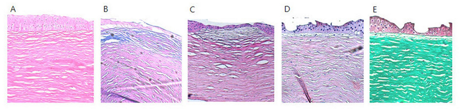
 Figure 3 of
Park, Mol Vis 2015; 21:1201-1209.
Figure 3 of
Park, Mol Vis 2015; 21:1201-1209.