Figure 7. Collagen I protein levels are reduced in KC corneas. The representative photomicrographs show staining for COL I (Brown color)
with counterstain hematoxylin (blue color) for nuclei. The staining is observed in the corneal stroma, as indicated by the
arrows. Three independent representative samples for KC and control corneas are presented at 40X magnification. Scale bar:
10 µm.
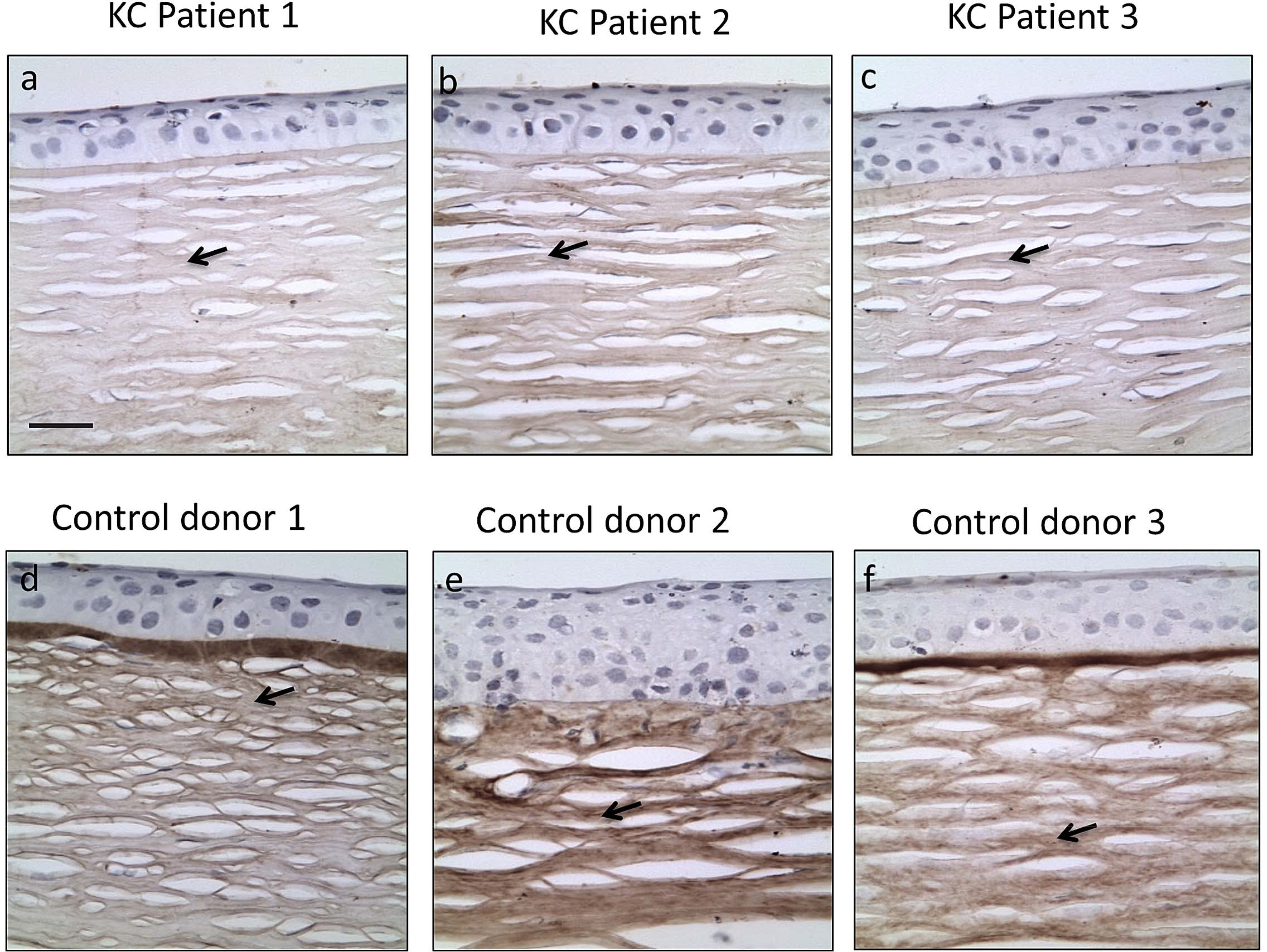
 Figure 7 of
Shetty, Mol Vis 2015; 21:12-25.
Figure 7 of
Shetty, Mol Vis 2015; 21:12-25.