Figure 6. Control staining for immunohistochemistry (IHC) experiments. Photomicrographs of control staining slides for IHC studies at
40X magnification; A-C: KC patient cornea; D-F: Control donor cornea. Actin staining (A and D); negative control without primary antibody, secondary antibody alone (B and E); hematoxylin and eosin (H & E) stain (C and F). Scale bar: 10 µm.
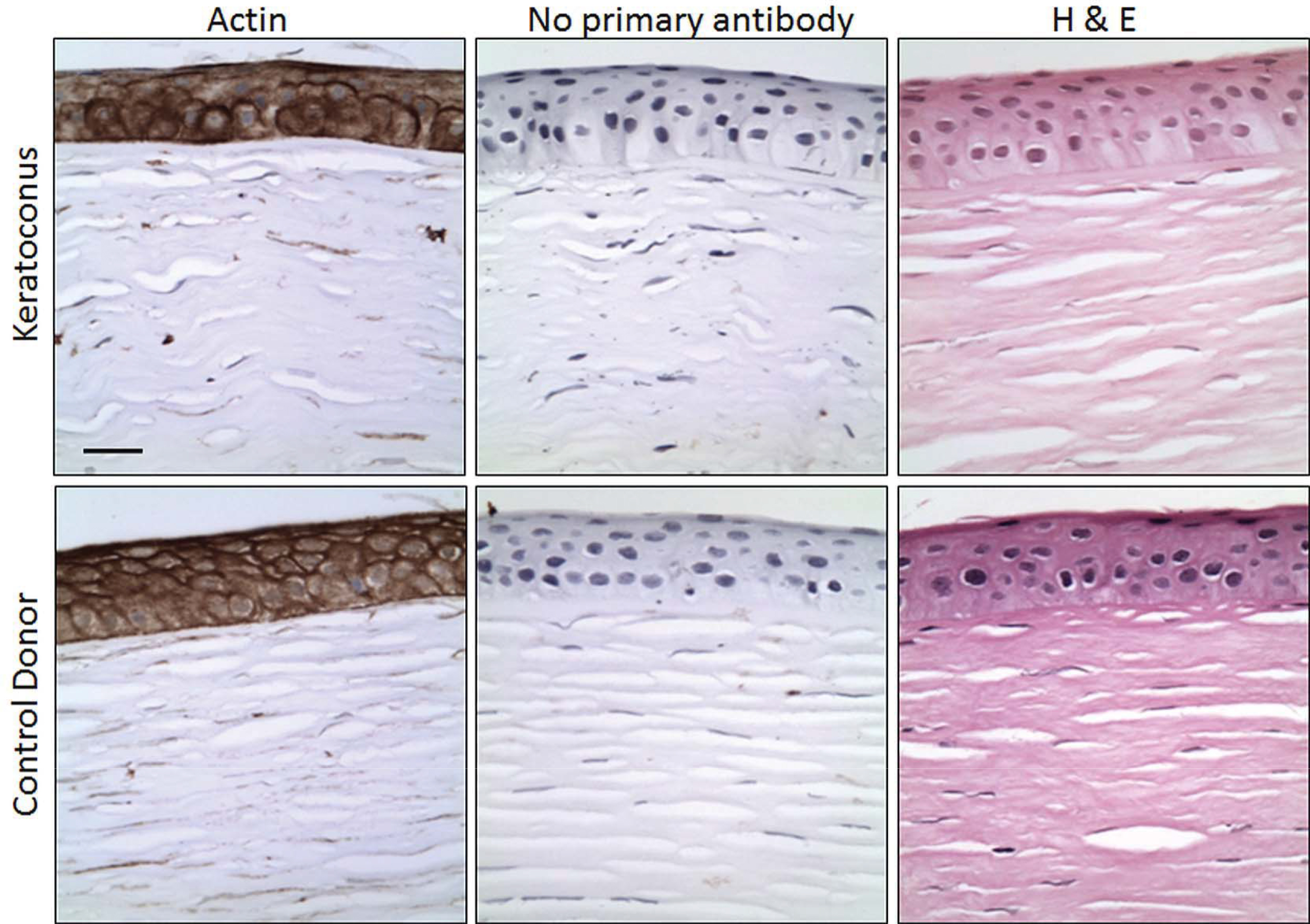
 Figure 6 of
Shetty, Mol Vis 2015; 21:12-25.
Figure 6 of
Shetty, Mol Vis 2015; 21:12-25.