Figure 1. Characterization of human Tenon’s fibroblast by vimentin and DAPI staining and cells morphological changes due to ranibizumab
treatment. A: Cytoplasm stained in green (Vimentin). B: Nucleus stained in blue (DAPI). C: Merge. D: Monochrome. E: Untreated HTF. F: HTF treated with Ranibizumab 0.5 mg/ml.
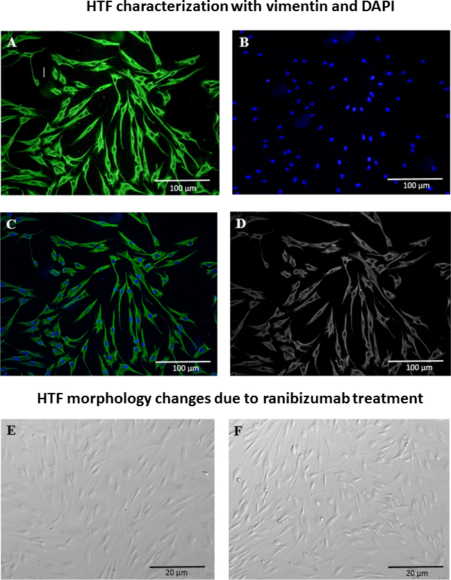
 Figure 1 of
Noh, Mol Vis 2015; 21:1191-1200.
Figure 1 of
Noh, Mol Vis 2015; 21:1191-1200.