Figure 3. Effects of anti-miR-155 on retinal neovascularization in the OIR model. P7 mice were exposed to hyperoxia for five days. On
P12, the pups were intravitreally injected with 1.5 μl of anti-miR-155. On P18, the pups were euthanized to detect the retinal
non-vascularization areas and neovascular numbers. A and B show representative images of FITC-dextran-stained retinas from various treatment groups and the results of statistical analysis.
C and D show representative mouse retina sections of the nuclei of neovascularization extending beyond the inner limiting membrane
(ILM) into the vitreous body of the anti-ctrl and anti-miR-155 treated groups and the results of statistical analysis. The
data are presented as mean ± SD. *p<0.05, **p<0.01, ***p<0.001. Scale bar: 50 μm.
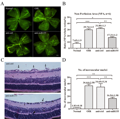
 Figure 3 of
Zhuang, Mol Vis 2015; 21:1173-1184.
Figure 3 of
Zhuang, Mol Vis 2015; 21:1173-1184.