Figure 2. NKCC1 and KCC2b in situ immunofluorescence detection in young adult rabbit lens. A, B: NKCC1 mAb and anti-KCC2 antibody detection in young adult rabbit lens (100X). C–E: NKCC1 expression in the cortical fiber cells is decreased as fiber cells mature in the lens interior. KCC2 is detected in
fiber cells closer to the lens center. Merged images C, D are shown in panel E (200X). F: KCC2b is detected at the fiber cell membrane borders in mature interior fiber cells (200X). G: KCC2b viewed at 600X magnification. Rabbit fiber cells are about 10 μm across in their longest dimension. The asterisks
identify comparable positions in the panels.
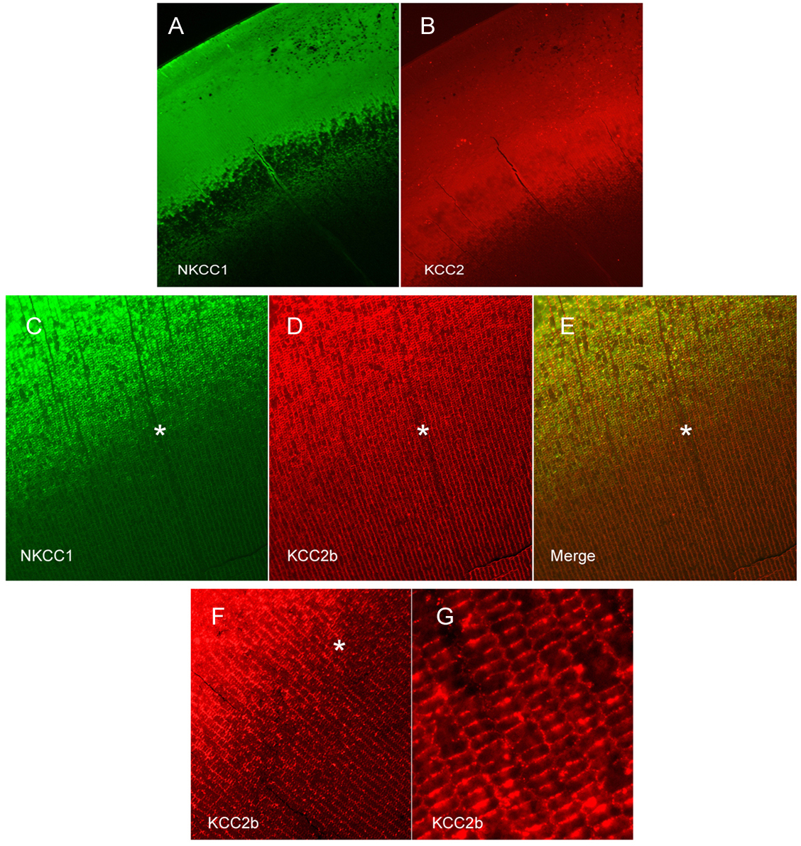
 Figure 2 of
Frederikse, Mol Vis 2015; 21:1142-1150.
Figure 2 of
Frederikse, Mol Vis 2015; 21:1142-1150.