Figure 2. Evaluation of conjunctival epithelial cell sheets fabricated with a temperature-responsive culture dish. A: Phase contrast microscopy showed that after culture for 4 days, the rabbit conjunctival epithelial cells (rCjECs) reached
confluence. PAS staining in conjunctival epithelial cell sheets. The positive cells indicate the presence of goblet cells
(arrowhead indicated goblet cells). B: Harvest of a conjunctival epithelial cell sheet. *** represents the cell sheets, * represents the culture surface (C). D: Hematoxylin and eosin (H&E) staining in conjunctival epithelial cell sheets. The result showed 4 to 5 cell layers. E, F: Immunohistochemical analysis of the cell sheets. CK4, CK19 staining for non-goblet cells. G: MUC5ac staining for goblet cells. Nuclei were counterstained with 4',6-diamidino-2-phenylindole dihydrochloride (DAPI).
Scale bars: 200 μm in A, B, C, D; 100 μm in E, F, G.
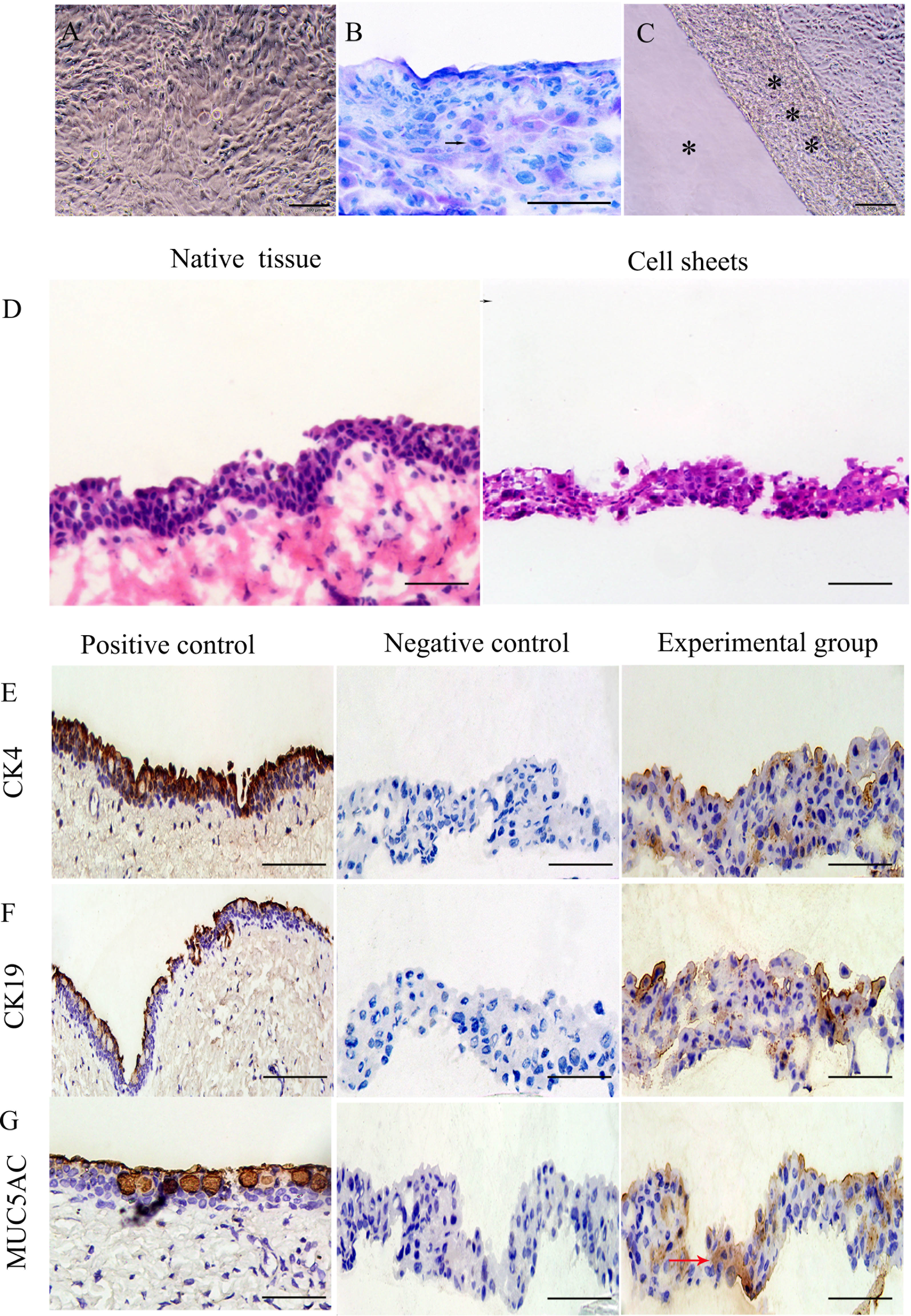
 Figure 2 of
Yao, Mol Vis 2015; 21:1113-1121.
Figure 2 of
Yao, Mol Vis 2015; 21:1113-1121.