Figure 1. The clinical retinal phenotype of two C3H congenic strains wild-type for Pde6b (used as the sighted control strains) and four C3H rd1 substrains. A: Normal fundus. B: Normal retinal structure by image-guided optical coherence tomography (OCT) at 6 weeks of age (representative strain is
C3A.BLiA-Pde6b+/J). C: Retinal vessels are attenuated, and areas of retinal depigmentation are observed. D: Retinal degeneration with image-guided OCT at 6 weeks of age (the representative strain is C3H/HeJ). The green line across
the fundus image indicates the location of the B-scan.
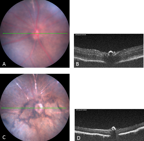
 Figure 1 of
Chang, Mol Vis 2015; 21:1101-1105.
Figure 1 of
Chang, Mol Vis 2015; 21:1101-1105.