Figure 7. Cfh localization within rod and cone photoreceptors. A sagittal section from a male BALB/c mouse eye immunolabeled for rhodopsin
(green), Cfh (red), and a 4',6-diamidino-2-phenylindole (DAPI) nuclear stain (blue) showed only a localized band of Cfh in
the central region of the OS (white arrow), but not throughout the OS. In addition, occasional Cfh labeling of cells believed
to be cone cell IS was observed (white arrowheads; A). A sagittal section from a male BALB/c mouse eye immunolabeled for blue and red/green opsins (white), Cfh (red), and a DAPI
nuclear stain (blue) showed a similar band of Cfh in the central region of the OS (green arrow; B). Cone cells labeled with the opsin antibodies (green arrowheads) exhibit a polarized distribution of blue and red/green
opsins (white) and Cfh (red). A coronal section from a male C57BL/6 eye double labeled with fluorescein conjugated WGA (green),
which labels the matrix domains surrounding rod photoreceptors, and Cfh (red) showing Cfh within rod photoreceptor OS (C). The delineated area of interest is magnified and shown on the right. Cfh is clearly localized within the rods (arrows;
D). A coronal section from a male C57BL/6 eye labeled with fluorescein conjugated PNA (green), which binds cone matrix domains,
and Cfh (red) showing Cfh is clearly localized within the cone cells (arrows; E). The delineated area of interest is magnified and shown on the right (F). IS=photoreceptor inner segments; OS=outer segments; RPE=retinal pigment epithelium; WGA=wheat germ agglutinin; PNA=peanut
agglutinin. Scale bar=20 μm.
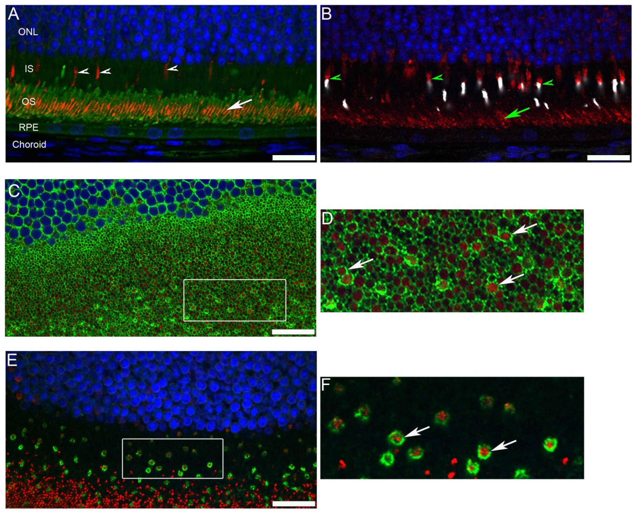
 Figure 7 of
Smit-McBride, Mol Vis 2015; 21:110-123.
Figure 7 of
Smit-McBride, Mol Vis 2015; 21:110-123.