Figure 6. Immunohistochemistry for Cfh protein expression in the BALB/c, 129/Sv, and 129/Sv Cfh−/− eye. A BALB/c male eye sectioned near the optic nerve labeled for Cfh using two anti-Cfh antibodies (C, D), along with its paired negative control (A, B). Goat anti-mouse Cfh antibody predominately labeled photoreceptor outer segments (OS) and what appear to be cone cell photoreceptor
(arrow heads) inner segments (IS; C). The rabbit anti-mouse Cfh antibody labeled OS, the apical edge of the retinal pigment epithelium (RPE) cells, and only
lightly labeled the RPE cytoplasm (D). The 129/Sv background strain eyes and Cfh knockout (129/Sv Cfh −/−) eyes paired with their negative controls (E, F, I, J) immunolabeled with goat anti-mouse Cfh antibody (G, H) or rabbit anti-mouse Cfh antibody (K, L). Cfh labeling was observed in the Cfh knockout eyes (H, L) using both antibodies, but the signal was greatly reduced compared to the wild-type (WT) eyes (G, K). The residual presence of the signal could be due to cross-reactivity with Cfhr2 protein, since both antibodies recognize
Cfh and Cfhr2 proteins. Scale bar=20 μm.
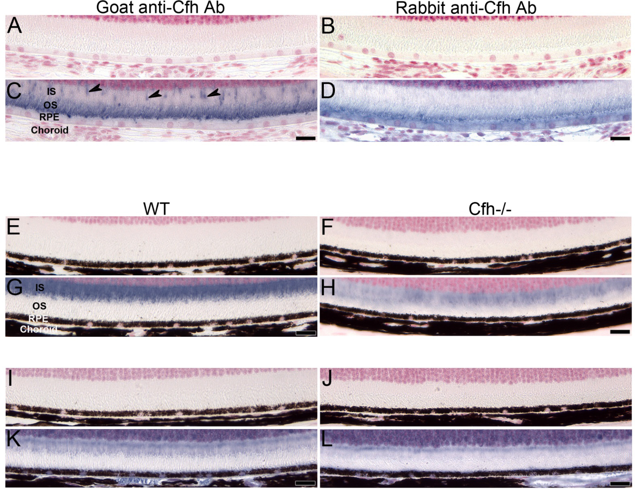
 Figure 6 of
Smit-McBride, Mol Vis 2015; 21:110-123.
Figure 6 of
Smit-McBride, Mol Vis 2015; 21:110-123.